5JXE | pdb_00005jxe

Human PD-1 ectodomain complexed with Pembrolizumab Fab


Experimental Data Snapshot

  • Method: X-RAY DIFFRACTION
  • Resolution: 2.90 Å
  • R-Value Free: 
    0.286 (Depositor), 0.288 (DCC) 
  • R-Value Work: 
    0.261 (Depositor), 0.262 (DCC) 
  • R-Value Observed: 
    0.262 (Depositor) 

Starting Models: experimental
View more details

wwPDB Validation 3D Report Full Report

Validation slider image for 5JXE

This is version 1.4 of the entry. See complete history

Literature

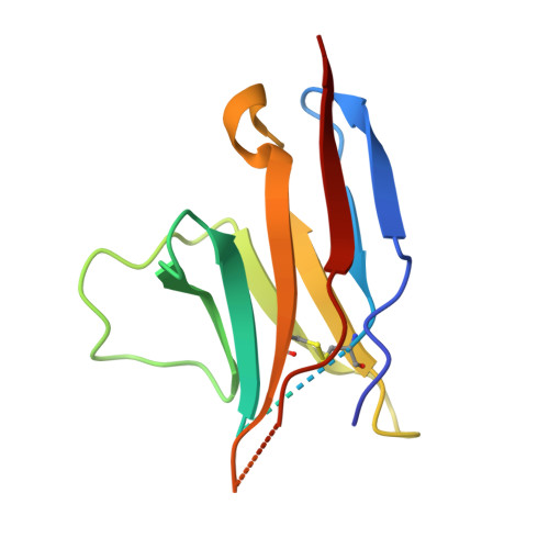
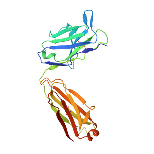
Structural basis for blocking PD-1-mediated immune suppression by therapeutic antibody pembrolizumab.

Na, Z.Yeo, S.P.Bharath, S.R.Bowler, M.W.Balijkcij, E.Wang, C.I.Song, H.

(2017) Cell Res 27: 147-150

  • DOI: https://doi.org/10.1038/cr.2016.77
  • Primary Citation Related Structures: 
    5JXE

  • Organizational Affiliation
    • Institute of Molecular and Cell Biology, 61 Biopolis Drive, Singapore 138673, Singapore.

Macromolecule Content 

  • Total Structure Weight: 120.33 kDa 
  • Atom Count: 7,851 
  • Modeled Residue Count: 1,043 
  • Deposited Residue Count: 1,100 
  • Unique protein chains: 3

Macromolecules

Find similar proteins by:|  3D Structure
Entity ID: 1
MoleculeChains  Sequence LengthOrganismDetailsImage
Programmed cell death protein 1
A, B
114Homo sapiensMutation(s): 1 
Gene Names: PDCD1PD1
UniProt & NIH Common Fund Data Resources
Find proteins for Q15116 (Homo sapiens)
Explore Q15116 
Go to UniProtKB:  Q15116
PHAROS:  Q15116
GTEx:  ENSG00000188389 
Entity Groups
Sequence Clusters30% Identity50% Identity70% Identity90% Identity95% Identity100% Identity
UniProt GroupQ15116
Sequence Annotations
Expand
Reference Sequence
Find similar proteins by:|  3D Structure
Entity ID: 2
MoleculeChains  Sequence LengthOrganismDetailsImage
Pembrolizumab Fab light chainC,
E [auth F]
218Homo sapiensMutation(s): 0 
Find similar proteins by:|  3D Structure
Entity ID: 3
MoleculeChains  Sequence LengthOrganismDetailsImage
Pembrolizumab Fab heavy chainD,
F [auth G]
218Homo sapiensMutation(s): 0 

Experimental Data & Validation

Experimental Data

  • Method: X-RAY DIFFRACTION
  • Resolution: 2.90 Å
  • R-Value Free:  0.286 (Depositor), 0.288 (DCC) 
  • R-Value Work:  0.261 (Depositor), 0.262 (DCC) 
  • R-Value Observed: 0.262 (Depositor) 
Space Group: P 65 2 2
Unit Cell:
Length ( Å )Angle ( ˚ )
a = 105.554α = 90
b = 105.554β = 90
c = 380.048γ = 120
Software Package:
Software NamePurpose
PHENIXrefinement
XDSdata reduction
Aimlessdata scaling
PHASERphasing

Structure Validation

View Full Validation Report


Entry History 

Deposition Data

Revision History  (Full details and data files)

  • Version 1.0: 2016-08-10
    Type: Initial release
  • Version 1.1: 2016-09-07
    Changes: Experimental preparation
  • Version 1.2: 2017-12-06
    Changes: Database references, Derived calculations
  • Version 1.3: 2023-11-08
    Changes: Data collection, Database references, Refinement description
  • Version 1.4: 2024-10-23
    Changes: Structure summary