5JWO

Crystal structure of foldswitch-stabilized KaiB in complex with the N-terminal CI domain of KaiC from Thermosynechococcus elongatus


Experimental Data Snapshot

  • Method: X-RAY DIFFRACTION
  • Resolution: 1.80 Å
  • R-Value Free: 0.257 
  • R-Value Work: 0.219 
  • R-Value Observed: 0.221 

wwPDB Validation   3D Report Full Report


Ligand Structure Quality Assessment 


This is version 1.3 of the entry. See complete history


Literature

Structural basis of the day-night transition in a bacterial circadian clock.

Tseng, R.Goularte, N.F.Chavan, A.Luu, J.Cohen, S.E.Chang, Y.G.Heisler, J.Li, S.Michael, A.K.Tripathi, S.Golden, S.S.LiWang, A.Partch, C.L.

(2017) Science 355: 1174-1180

  • DOI: https://doi.org/10.1126/science.aag2516
  • Primary Citation of Related Structures:  
    5JWO, 5JWQ, 5JWR, 5JYT, 5JYU, 5JYV

  • PubMed Abstract: 

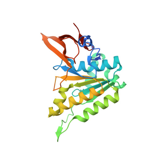
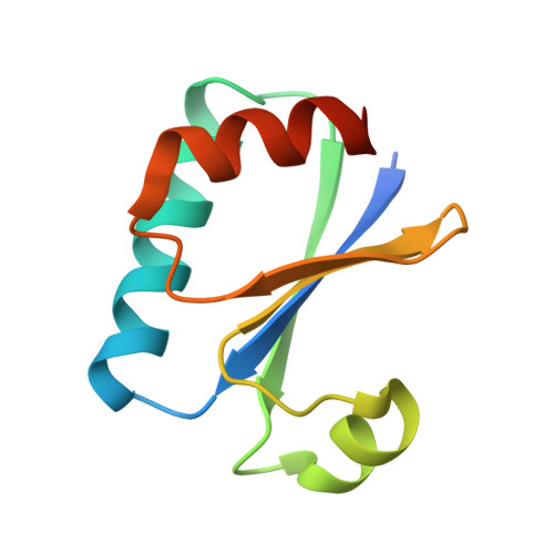
    Circadian clocks are ubiquitous timing systems that induce rhythms of biological activities in synchrony with night and day. In cyanobacteria, timing is generated by a posttranslational clock consisting of KaiA, KaiB, and KaiC proteins and a set of output signaling proteins, SasA and CikA, which transduce this rhythm to control gene expression. Here, we describe crystal and nuclear magnetic resonance structures of KaiB-KaiC,KaiA-KaiB-KaiC, and CikA-KaiB complexes. They reveal how the metamorphic properties of KaiB, a protein that adopts two distinct folds, and the post-adenosine triphosphate hydrolysis state of KaiC create a hub around which nighttime signaling events revolve, including inactivation of KaiA and reciprocal regulation of the mutually antagonistic signaling proteins, SasA and CikA.


  • Organizational Affiliation

    Quantitative and Systems Biology, University of California, Merced, CA 95343, USA.


Macromolecules
Find similar proteins by:  (by identity cutoff)  |  3D Structure
Entity ID: 1
MoleculeChains Sequence LengthOrganismDetailsImage
Circadian clock protein kinase KaiC247Thermosynechococcus vestitus BP-1Mutation(s): 2 
Gene Names: kaiCtlr0483
EC: 2.7.11.1
UniProt
Find proteins for Q79V60 (Thermosynechococcus vestitus (strain NIES-2133 / IAM M-273 / BP-1))
Explore Q79V60 
Go to UniProtKB:  Q79V60
Entity Groups  
Sequence Clusters30% Identity50% Identity70% Identity90% Identity95% Identity100% Identity
UniProt GroupQ79V60
Sequence Annotations
Expand
  • Reference Sequence
Find similar proteins by:  (by identity cutoff)  |  3D Structure
Entity ID: 2
MoleculeChains Sequence LengthOrganismDetailsImage
Circadian clock protein KaiB99Thermosynechococcus vestitus BP-1Mutation(s): 4 
Gene Names: kaiBtlr0482
UniProt
Find proteins for Q79V61 (Thermosynechococcus vestitus (strain NIES-2133 / IAM M-273 / BP-1))
Explore Q79V61 
Go to UniProtKB:  Q79V61
Entity Groups  
Sequence Clusters30% Identity50% Identity70% Identity90% Identity95% Identity100% Identity
UniProt GroupQ79V61
Sequence Annotations
Expand
  • Reference Sequence
Small Molecules
Ligands 1 Unique
IDChains Name / Formula / InChI Key2D Diagram3D Interactions
ADP
Query on ADP

Download Ideal Coordinates CCD File 
C [auth A]ADENOSINE-5'-DIPHOSPHATE
C10 H15 N5 O10 P2
XTWYTFMLZFPYCI-KQYNXXCUSA-N
Experimental Data & Validation

Experimental Data

  • Method: X-RAY DIFFRACTION
  • Resolution: 1.80 Å
  • R-Value Free: 0.257 
  • R-Value Work: 0.219 
  • R-Value Observed: 0.221 
  • Space Group: P 2 21 21
Unit Cell:
Length ( Å )Angle ( ˚ )
a = 42.48α = 90
b = 56.59β = 90
c = 146.14γ = 90
Software Package:
Software NamePurpose
PHENIXrefinement
iMOSFLMdata reduction
SCALAdata scaling
PHASERphasing

Structure Validation

View Full Validation Report


Ligand Structure Quality Assessment 


Entry History & Funding Information

Deposition Data


Funding OrganizationLocationGrant Number
National Institutes of Health/National Institute of General Medical Sciences (NIH/NIGMS)United StatesGM107521
National Institutes of Health/National Institute of General Medical Sciences (NIH/NIGMS)United StatesGM107069

Revision History  (Full details and data files)

  • Version 1.0: 2017-03-29
    Type: Initial release
  • Version 1.1: 2017-09-20
    Changes: Author supporting evidence
  • Version 1.2: 2019-12-25
    Changes: Author supporting evidence
  • Version 1.3: 2024-03-06
    Changes: Data collection, Database references