5JWB | pdb_00005jwb

Structure of NDH2 from plasmodium falciparum in complex with NADH


Experimental Data Snapshot

  • Method: X-RAY DIFFRACTION
  • Resolution: 2.70 Å
  • R-Value Free: 
    0.226 (Depositor), 0.207 (DCC) 
  • R-Value Work: 
    0.180 (Depositor) 
  • R-Value Observed: 
    0.183 (Depositor) 

Starting Model: experimental
View more details

wwPDB Validation 3D Report Full Report

Validation slider image for 5JWB

Ligand Structure Quality Assessment 


This is version 1.2 of the entry. See complete history

Literature

Target Elucidation by Cocrystal Structures of NADH-Ubiquinone Oxidoreductase of Plasmodium falciparum (PfNDH2) with Small Molecule To Eliminate Drug-Resistant Malaria

Yang, Y.Yu, Y.Li, X.Li, J.Wu, Y.Yu, J.Ge, J.Huang, Z.Jiang, L.Rao, Y.Yang, M.

(2017) J Med Chem 60: 1994-2005

  • DOI: https://doi.org/10.1021/acs.jmedchem.6b01733
  • Primary Citation Related Structures: 
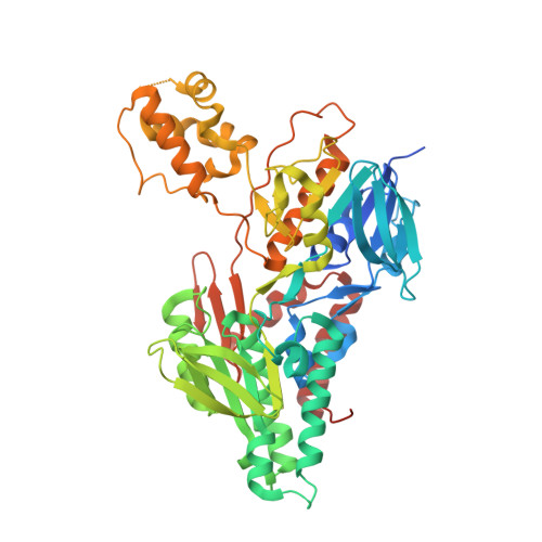
    5JWA, 5JWB, 5JWC

  • PubMed Abstract: 

    Drug-resistant malarial strains have been continuously emerging recently, which posts a great challenge for the global health. Therefore, new antimalarial drugs with novel targeting mechanisms are urgently needed for fighting drug-resistant malaria. NADH-ubiquinone oxidoreductase of Plasmodium falciparum (PfNDH2) represents a viable target for antimalarial drug development. However, the absence of structural information on PfNDH2 limited rational drug design and further development. Herein, we report high resolution crystal structures of the PfNDH2 protein for the first time in Apo-, NADH-, and RYL-552 (a new inhibitor)-bound states. The PfNDH2 inhibitor exhibits excellent potency against both drug-resistant strains in vitro and parasite-infected mice in vivo via a potential allosteric mechanism. Furthermore, it was found that the inhibitor can be used in combination with dihydroartemisinin (DHA) synergistically. These findings not only are important for malarial PfNDH2 protein-based drug development but could also have broad implications for other NDH2-containing pathogenic microorganisms such as Mycobacterium tuberculosis.


  • Organizational Affiliation
    • MOE Key Laboratory of Protein Sciences, School of Pharmaceutical Sciences, Tsinghua University , Beijing 100084, China.

Macromolecule Content 

  • Total Structure Weight: 126.66 kDa 
  • Atom Count: 8,456 
  • Modeled Residue Count: 985 
  • Deposited Residue Count: 1,042 
  • Unique protein chains: 1

Macromolecules

Find similar proteins by:|  3D Structure
Entity ID: 1
MoleculeChains  Sequence LengthOrganismDetailsImage
Type II NADH:ubiquinone oxidoreductaseA,
B [auth H]
521Plasmodium falciparum 3D7Mutation(s): 0 
Gene Names: PF3D7_0915000
EC: 1.6.5.3 (PDB Primary Data), 1.6.5.9 (UniProt)
UniProt
Find proteins for Q8I302 (Plasmodium falciparum (isolate 3D7))
Explore Q8I302 
Go to UniProtKB:  Q8I302
Entity Groups
Sequence Clusters30% Identity50% Identity70% Identity90% Identity95% Identity100% Identity
UniProt GroupQ8I302
Sequence Annotations
Expand
Reference Sequence

Small Molecules

Ligands 6 Unique
IDChains Name / Formula / InChI Key2D Diagram3D Interactions
FAD

Query on FAD


Download:Ideal Coordinates CCD File
C [auth A],
R [auth H]
FLAVIN-ADENINE DINUCLEOTIDE
C27 H33 N9 O15 P2
VWWQXMAJTJZDQX-UYBVJOGSSA-N
NAD

Query on NAD


Download:Ideal Coordinates CCD File
J [auth A],
W [auth H]
NICOTINAMIDE-ADENINE-DINUCLEOTIDE
C21 H27 N7 O14 P2
BAWFJGJZGIEFAR-NNYOXOHSSA-N
TRT

Query on TRT


Download:Ideal Coordinates CCD File
H [auth A]
I [auth A]
K [auth A]
Q [auth H]
X [auth H]
H [auth A],
I [auth A],
K [auth A],
Q [auth H],
X [auth H],
Y [auth H]
FRAGMENT OF TRITON X-100
C21 H36 O4
HEUDUECKTWTQQR-UHFFFAOYSA-N
CXS

Query on CXS


Download:Ideal Coordinates CCD File
CA [auth H],
P [auth A]
3-CYCLOHEXYL-1-PROPYLSULFONIC ACID
C9 H19 N O3 S
PJWWRFATQTVXHA-UHFFFAOYSA-N
ACT

Query on ACT


Download:Ideal Coordinates CCD File
AA [auth H]
BA [auth H]
L [auth A]
M [auth A]
N [auth A]
AA [auth H],
BA [auth H],
L [auth A],
M [auth A],
N [auth A],
O [auth A],
Z [auth H]
ACETATE ION
C2 H3 O2
QTBSBXVTEAMEQO-UHFFFAOYSA-M
MG

Query on MG


Download:Ideal Coordinates CCD File
D [auth A]
E [auth A]
F [auth A]
G [auth A]
S [auth H]
D [auth A],
E [auth A],
F [auth A],
G [auth A],
S [auth H],
T [auth H],
U [auth H],
V [auth H]
MAGNESIUM ION
Mg
JLVVSXFLKOJNIY-UHFFFAOYSA-N

Experimental Data & Validation

Experimental Data

  • Method: X-RAY DIFFRACTION
  • Resolution: 2.70 Å
  • R-Value Free:  0.226 (Depositor), 0.207 (DCC) 
  • R-Value Work:  0.180 (Depositor) 
  • R-Value Observed: 0.183 (Depositor) 
Space Group: I 41
Unit Cell:
Length ( Å )Angle ( ˚ )
a = 191.53α = 90
b = 191.53β = 90
c = 91.258γ = 90
Software Package:
Software NamePurpose
PHENIXrefinement
SCALEPACKdata scaling
PDB_EXTRACTdata extraction
HKL-2000data reduction
PHASERphasing

Structure Validation

View Full Validation Report


Ligand Structure Quality Assessment 


Entry History 

Deposition Data

Revision History  (Full details and data files)

  • Version 1.0: 2017-03-29
    Type: Initial release
  • Version 1.1: 2019-12-25
    Changes: Data collection
  • Version 1.2: 2023-11-08
    Changes: Data collection, Database references, Derived calculations, Refinement description