


Structure of F420 binding protein, MSMEG_6526, from Mycobacterium smegmatis with F420 bound
Lee, B.M., Carr, P.D., Jackson, C.J.To be published.
Experimental Data Snapshot
Starting Model: experimental
View more details
Entity ID: 1 | |||||
|---|---|---|---|---|---|
| Molecule | Chains | Sequence Length | Organism | Details | Image |
| Pyridoxamine 5'-phosphate oxidase-like FMN-binding protein | 154 | Mycolicibacterium smegmatis | Mutation(s): 0 Gene Names: ERS451418_06325 | 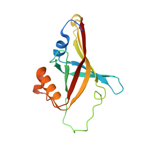 | |
UniProt | |||||
Entity Groups | |||||
| Sequence Clusters | 30% Identity50% Identity70% Identity90% Identity95% Identity100% Identity | ||||
| UniProt Group | A0R6F1 | ||||
Sequence AnnotationsExpand | |||||
Reference Sequence | |||||
| Ligands 4 Unique | |||||
|---|---|---|---|---|---|
| ID | Chains | Name / Formula / InChI Key | 2D Diagram | 3D Interactions | |
| F42 Download:Ideal Coordinates CCD File | G [auth A] I [auth B] J [auth B] N [auth C] R [auth D] | COENZYME F420 C29 H36 N5 O18 P GEHSZWRGPHDXJO-NALJQGANSA-N |  | ||
| MRD Download:Ideal Coordinates CCD File | M [auth B], P [auth C] | (4R)-2-METHYLPENTANE-2,4-DIOL C6 H14 O2 SVTBMSDMJJWYQN-RXMQYKEDSA-N |  | ||
| MPD Download:Ideal Coordinates CCD File | H [auth A] K [auth B] L [auth B] O [auth C] S [auth E] | (4S)-2-METHYL-2,4-PENTANEDIOL C6 H14 O2 SVTBMSDMJJWYQN-YFKPBYRVSA-N |  | ||
| NA Download:Ideal Coordinates CCD File | Q [auth D] | SODIUM ION Na FKNQFGJONOIPTF-UHFFFAOYSA-N |  | ||
| Length ( Å ) | Angle ( ˚ ) |
|---|---|
| a = 139.951 | α = 90 |
| b = 84.241 | β = 90.85 |
| c = 75.98 | γ = 90 |
| Software Name | Purpose |
|---|---|
| REFMAC | refinement |
| iMOSFLM | data reduction |
| Aimless | data scaling |
| MOLREP | phasing |


