5JTV

USP7CD-UBL45 in complex with Ubiquitin


Experimental Data Snapshot

  • Method: X-RAY DIFFRACTION
  • Resolution: 3.31 Å
  • R-Value Free: 0.269 
  • R-Value Work: 0.239 
  • R-Value Observed: 0.240 

wwPDB Validation   3D Report Full Report


This is version 1.1 of the entry. See complete history


Literature

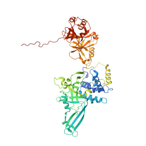
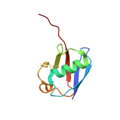
Molecular Understanding of USP7 Substrate Recognition and C-Terminal Activation.

Rouge, L.Bainbridge, T.W.Kwok, M.Tong, R.Di Lello, P.Wertz, I.E.Maurer, T.Ernst, J.A.Murray, J.

(2016) Structure 24: 1335-1345

  • DOI: https://doi.org/10.1016/j.str.2016.05.020
  • Primary Citation of Related Structures:  
    5J7T, 5JTJ, 5JTV

  • PubMed Abstract: 

    The deubiquitinating enzyme USP7 has a pivotal role in regulating the stability of proteins involved in fundamental cellular processes of normal biology and disease. Despite the importance of USP7, the mechanisms underlying substrate recognition and catalytic activation are poorly understood. Here we present structural, biochemical, and biophysical analyses elucidating the molecular mechanism by which the C-terminal 19 amino acids of USP7 (residues 1084-1102) enhance the ubiquitin cleavage activity of the deubiquitinase (DUB) domain. Our data demonstrate that the C-terminal peptide binds the activation cleft in the catalytic domain and stabilizes the catalytically competent conformation of USP7. Additional structures of longer fragments of USP7, as well as solution studies, provide insight into full-length USP7, the role of the UBL domains, and demonstrate that both substrate recognition and deubiquitinase activity are highly regulated by the catalytic and noncatalytic domains of USP7, a feature that could be essential for the proper function of multi-domain DUBs.


  • Organizational Affiliation

    Department of Structural Biology, Genentech, South San Francisco, CA 94080, USA.


Macromolecules
Find similar proteins by:  (by identity cutoff)  |  3D Structure
Entity ID: 1
MoleculeChains Sequence LengthOrganismDetailsImage
Ubiquitin carboxyl-terminal hydrolase 7
A, C, E, G
594Homo sapiensMutation(s): 0 
Gene Names: USP7HAUSP
EC: 3.4.19.12
UniProt & NIH Common Fund Data Resources
Find proteins for Q93009 (Homo sapiens)
Explore Q93009 
Go to UniProtKB:  Q93009
PHAROS:  Q93009
GTEx:  ENSG00000187555 
Entity Groups  
Sequence Clusters30% Identity50% Identity70% Identity90% Identity95% Identity100% Identity
UniProt GroupQ93009
Sequence Annotations
Expand
  • Reference Sequence
Find similar proteins by:  (by identity cutoff)  |  3D Structure
Entity ID: 2
MoleculeChains Sequence LengthOrganismDetailsImage
Polyubiquitin-B
B, D, F, H
76Homo sapiensMutation(s): 0 
Gene Names: UBB
UniProt & NIH Common Fund Data Resources
Find proteins for P0CG47 (Homo sapiens)
Explore P0CG47 
Go to UniProtKB:  P0CG47
PHAROS:  P0CG47
GTEx:  ENSG00000170315 
Entity Groups  
Sequence Clusters30% Identity50% Identity70% Identity90% Identity95% Identity100% Identity
UniProt GroupP0CG47
Sequence Annotations
Expand
  • Reference Sequence
Experimental Data & Validation

Experimental Data

  • Method: X-RAY DIFFRACTION
  • Resolution: 3.31 Å
  • R-Value Free: 0.269 
  • R-Value Work: 0.239 
  • R-Value Observed: 0.240 
  • Space Group: P 21 21 21
Unit Cell:
Length ( Å )Angle ( ˚ )
a = 113.611α = 90
b = 115.52β = 90
c = 257.822γ = 90
Software Package:
Software NamePurpose
PHENIXrefinement
DENZOdata reduction
SCALEPACKdata scaling
PHASERphasing
PDB_EXTRACTdata extraction

Structure Validation

View Full Validation Report


Entry History 

Deposition Data

Revision History  (Full details and data files)

  • Version 1.0: 2016-10-12
    Type: Initial release
  • Version 1.1: 2023-09-27
    Changes: Data collection, Database references, Derived calculations, Refinement description