Crystal structure of 6aJL2-R24G light chain variable domain: Does crystal packing explain amyloid fibril formation?
Rudino-Pinera, E., Pelaez-Aguilar, A.E., Amero, C., Diaz-Vilchis, A.(2019) Biochem Biophys Rep 20: 100682-100682
- PubMed: 31517067 Search on PubMedSearch on PubMed Central
- DOI: https://doi.org/10.1016/j.bbrep.2019.100682
- Primary Citation Related Structures:
5JPJ - PubMed Abstract:
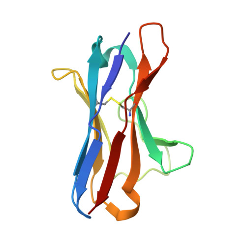
Light chain amyloidosis is one of the most common systemic amyloidosis, characterized by the deposition of immunoglobulin light variable domain as insoluble amyloid fibrils in vital organs, leading to the death of patients. Germline λ6a is closely related with this disease and has been reported that 25% of proteins encoded by this germline have a change at position 24 where an Arg is replaced by a Gly (R24G). This germline variant reduces protein stability and increases the propensity to form amyloid fibrils. In this work, the crystal structure of 6aJL2-R24G has been determined to 2.0 Å resolution by molecular replacement. Crystal belongs to space group I2 1 2 1 2 1 (PDB ID 5JPJ) and there are two molecules in the asymmetric unit. This 6aJL2-R24G structure as several related in PDB (PDB entries: 5C9K, 2W0K, 5IR3 and 1PW3) presents by crystal packing the formation of an octameric assembly in a helicoidal arrangement, which has been proposed as an important early stage in amyloid fibril aggregation. However, other structures of other protein variants in PDB (PDB entries: 3B5G, 3BDX, 2W0L, 1CD0 and 2CD0) do not make the octameric assembly, regardless their capacity to form fibers in vitro or in vivo . The analysis presented here shows that the ability to form the octameric assembly in a helicoidal arrangement in crystallized light chain immunoglobulin proteins is not required for amyloid fibril formation in vitro . In addition, the fundamental role of partially folded states in the amyloid fibril formation in vitro , is not described in any crystallographic structure published or analyzed here, being those structures, in any case examples of proteins in their native states. Those partially folded states have been recently described by cryo-EM studies, showing the necessity of structural changes in the variants before the amyloid fiber formation process starts.
- Departamento de Medicina Molecular y Bioprocesos, Instituto de Biotecnología, Universidad Nacional Autónoma de México, Avenida Universidad 2001, Colonia Chamilpa, Cuernavaca, Morelos, 62210, Mexico.
Organizational Affiliation: