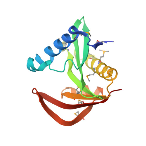
5JPH

Structure of a GNAT acetyltransferase SACOL1063 from Staphylococcus aureus in complex with CoA


Experimental Data Snapshot

  • Method: X-RAY DIFFRACTION
  • Resolution: 1.46 Å
  • R-Value Free: 0.170 
  • R-Value Work: 0.142 
  • R-Value Observed: 0.143 

wwPDB Validation   3D Report Full Report


Ligand Structure Quality Assessment 


This is version 1.4 of the entry. See complete history


Literature

Insight into the 3D structure and substrate specificity of previously uncharacterized GNAT superfamily acetyltransferases from pathogenic bacteria.

Majorek, K.A.Osinski, T.Tran, D.T.Revilla, A.Anderson, W.F.Minor, W.Kuhn, M.L.

(2016) Biochim Biophys Acta 1865: 55-64

  • DOI: https://doi.org/10.1016/j.bbapap.2016.10.011
  • Primary Citation of Related Structures:  
    5JPH, 5JQ4

  • PubMed Abstract: 

    Members of the Gcn5-related N-acetyltransferase (GNAT) superfamily catalyze the acetylation of a wide range of small molecule and protein substrates. Due to their abundance in all kingdoms of life and diversity of their functions, they are implicated in many aspects of eukaryotic and prokaryotic physiology. Although numerous GNATs have been identified thus far, many remain structurally and functionally uncharacterized. The elucidation of their structures and functions is critical for broadening our knowledge of this diverse and important superfamily. In this work, we present the structural and kinetic analyses of two previously uncharacterized bacterial acetyltransferases - SACOL1063 from Staphylococcus aureus strain COL and CD1211 from Clostridium difficile strain 630. Our structures of SACOL1063 show substantial flexibility of a loop that is likely responsible for substrate recognition and binding compared to structures of other homologs. In the CoA complex structure, we found two CoA molecules bound in both the canonical AcCoA/CoA-binding site and the acceptor-substrate-binding site. Our work also provides initial clues regarding the substrate specificity of these two enzymes; however, their native function(s) remain unknown. We found both proteins act as N- rather than O-acetyltransferases and preferentially acetylate l-threonine. The combination of structural and kinetic analyses of these two previously uncharacterized GNATs provides fundamental knowledge and a framework on which future studies can be built to elucidate their native functions.


  • Organizational Affiliation

    Department of Molecular Physiology and Biological Physics, University of Virginia, Charlottesville, VA 22908, USA; Center for Structural Genomics of Infectious Diseases (CSGID).


Macromolecules
Find similar proteins by:  (by identity cutoff)  |  3D Structure
Entity ID: 1
MoleculeChains Sequence LengthOrganismDetailsImage
Acetyltransferase SACOL1063
A, B, C
144Staphylococcus aureus subsp. aureus COLMutation(s): 0 
Gene Names: SACOL1063
EC: 2.3.1
UniProt
Find proteins for Q5HH30 (Staphylococcus aureus (strain COL))
Explore Q5HH30 
Go to UniProtKB:  Q5HH30
Entity Groups  
Sequence Clusters30% Identity50% Identity70% Identity90% Identity95% Identity100% Identity
UniProt GroupQ5HH30
Sequence Annotations
Expand
  • Reference Sequence
Small Molecules
Ligands 2 Unique
IDChains Name / Formula / InChI Key2D Diagram3D Interactions
COA
Query on COA

Download Ideal Coordinates CCD File 
D [auth A]
E [auth A]
G [auth B]
H [auth B]
J [auth C]
D [auth A],
E [auth A],
G [auth B],
H [auth B],
J [auth C],
K [auth C]
COENZYME A
C21 H36 N7 O16 P3 S
RGJOEKWQDUBAIZ-IBOSZNHHSA-N
CL
Query on CL

Download Ideal Coordinates CCD File 
F [auth A],
I [auth B],
L [auth C]
CHLORIDE ION
Cl
VEXZGXHMUGYJMC-UHFFFAOYSA-M
Modified Residues  1 Unique
IDChains TypeFormula2D DiagramParent
MSE
Query on MSE
A, B, C
L-PEPTIDE LINKINGC5 H11 N O2 SeMET
Experimental Data & Validation

Experimental Data

Unit Cell:
Length ( Å )Angle ( ˚ )
a = 50.088α = 90
b = 93.821β = 97.15
c = 68.91γ = 90
Software Package:
Software NamePurpose
HKL-2000data collection
HKL-2000data scaling
REFMACrefinement
PDB_EXTRACTdata extraction
HKL-3000data reduction
HKL-3000phasing

Structure Validation

View Full Validation Report


Ligand Structure Quality Assessment 


Entry History 

Deposition Data

Revision History  (Full details and data files)

  • Version 1.0: 2016-06-29
    Type: Initial release
  • Version 1.1: 2016-11-16
    Changes: Database references
  • Version 1.2: 2016-12-07
    Changes: Other
  • Version 1.3: 2022-04-13
    Changes: Database references, Structure summary
  • Version 1.4: 2024-11-13
    Changes: Data collection, Refinement description, Structure summary