5JLT

The crystal structure of the bacteriophage T4 MotA C-terminal domain in complex with dsDNA reveals a novel protein-DNA recognition motif


Experimental Data Snapshot

  • Method: X-RAY DIFFRACTION
  • Resolution: 2.96 Å
  • R-Value Free: 0.245 
  • R-Value Work: 0.220 
  • R-Value Observed: 0.221 

wwPDB Validation   3D Report Full Report


This is version 1.2 of the entry. See complete history


Literature

The phage T4 MotA transcription factor contains a novel DNA binding motif that specifically recognizes modified DNA.

Cuypers, M.G.Robertson, R.M.Knipling, L.Waddell, M.B.Moon, K.Hinton, D.M.White, S.W.

(2018) Nucleic Acids Res 46: 5308-5318

  • DOI: https://doi.org/10.1093/nar/gky292
  • Primary Citation of Related Structures:  
    5JLT

  • PubMed Abstract: 

    During infection, bacteriophage T4 produces the MotA transcription factor that redirects the host RNA polymerase to the expression of T4 middle genes. The C-terminal 'double-wing' domain of MotA binds specifically to the MotA box motif of middle T4 promoters. We report the crystal structure of this complex, which reveals a new mode of protein-DNA interaction. The domain binds DNA mostly via interactions with the DNA backbone, but the binding is enhanced in the specific cognate structure by additional interactions with the MotA box motif in both the major and minor grooves. The linker connecting the two MotA domains plays a key role in stabilizing the complex via minor groove interactions. The structure is consistent with our previous model derived from chemical cleavage experiments using the entire transcription complex. α- and β-d-glucosyl-5-hydroxymethyl-deoxycytosine replace cytosine in T4 DNA, and docking simulations indicate that a cavity in the cognate structure can accommodate the modified cytosine. Binding studies confirm that the modification significantly enhances the binding affinity of MotA for the DNA. Consequently, our work reveals how a DNA modification can extend the uniqueness of small DNA motifs to facilitate the specificity of protein-DNA interactions.


  • Organizational Affiliation

    Department of Structural Biology, St. Jude Children's Research Hospital, Memphis, TN 38105, USA.


Macromolecules

Find similar proteins by:  (by identity cutoff)  |  3D Structure
Entity ID: 1
MoleculeChains Sequence LengthOrganismDetailsImage
Middle transcription regulatory protein motA
A, B, C, D
125Tequatrovirus T4Mutation(s): 0 
Gene Names: motA
UniProt
Find proteins for P22915 (Enterobacteria phage T4)
Explore P22915 
Go to UniProtKB:  P22915
Entity Groups  
Sequence Clusters30% Identity50% Identity70% Identity90% Identity95% Identity100% Identity
UniProt GroupP22915
Sequence Annotations
Expand
  • Reference Sequence

Find similar nucleic acids by:  Sequence   |   3D Structure  

Entity ID: 2
MoleculeChains LengthOrganismImage
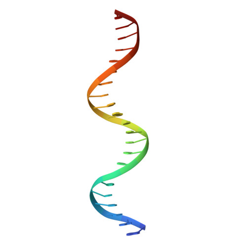
DNA (5'-D(*GP*AP*AP*GP*CP*TP*TP*TP*GP*CP*TP*TP*AP*AP*TP*AP*AP*TP*CP*CP*AP*C)-3')
E, H
22synthetic construct
Sequence Annotations
Expand
  • Reference Sequence

Find similar nucleic acids by:  Sequence   |   3D Structure  

Entity ID: 3
MoleculeChains LengthOrganismImage
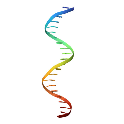
DNA (5'-D(*GP*TP*GP*GP*AP*TP*TP*AP*TP*TP*AP*AP*GP*CP*AP*AP*AP*GP*CP*TP*TP*C)-3')
F, G
22synthetic construct
Sequence Annotations
Expand
  • Reference Sequence
Experimental Data & Validation

Experimental Data

  • Method: X-RAY DIFFRACTION
  • Resolution: 2.96 Å
  • R-Value Free: 0.245 
  • R-Value Work: 0.220 
  • R-Value Observed: 0.221 
  • Space Group: P 61
  • Diffraction Data: https://doi.org/10.18430/m35jlt
Unit Cell:
Length ( Å )Angle ( ˚ )
a = 72.27α = 90
b = 72.27β = 90
c = 279.369γ = 120
Software Package:
Software NamePurpose
PHENIXrefinement
XDSdata reduction
SCALAdata scaling
PHASERphasing

Structure Validation

View Full Validation Report


Entry History 

Deposition Data

Revision History  (Full details and data files)

  • Version 1.0: 2017-05-03
    Type: Initial release
  • Version 1.1: 2019-11-27
    Changes: Database references
  • Version 1.2: 2023-09-27
    Changes: Data collection, Database references, Refinement description