5JLH

Cryo-EM structure of a human cytoplasmic actomyosin complex at near-atomic resolution


Experimental Data Snapshot

  • Method: ELECTRON MICROSCOPY
  • Resolution: 3.90 Å
  • Aggregation State: FILAMENT 
  • Reconstruction Method: SINGLE PARTICLE 

wwPDB Validation   3D Report Full Report


This is version 1.6 of the entry. See complete history


Literature

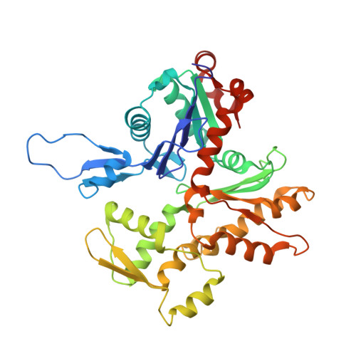
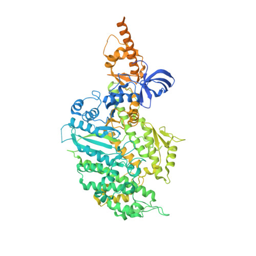
Cryo-EM structure of a human cytoplasmic actomyosin complex at near-atomic resolution.

Ecken, J.V.Heissler, S.M.Pathan-Chhatbar, S.Manstein, D.J.Raunser, S.

(2016) Nature 534: 724-728

  • DOI: https://doi.org/10.1038/nature18295
  • Primary Citation of Related Structures:  
    5JLF, 5JLH

  • PubMed Abstract: 

    The interaction of myosin with actin filaments is the central feature of muscle contraction and cargo movement along actin filaments of the cytoskeleton. The energy for these movements is generated during a complex mechanochemical reaction cycle. Crystal structures of myosin in different states have provided important structural insights into the myosin motor cycle when myosin is detached from F-actin. The difficulty of obtaining diffracting crystals, however, has prevented structure determination by crystallography of actomyosin complexes. Thus, although structural models exist of F-actin in complex with various myosins, a high-resolution structure of the F-actin–myosin complex is missing. Here, using electron cryomicroscopy, we present the structure of a human rigor actomyosin complex at an average resolution of 3.9 Å. The structure reveals details of the actomyosin interface, which is mainly stabilized by hydrophobic interactions. The negatively charged amino (N) terminus of actin interacts with a conserved basic motif in loop 2 of myosin, promoting cleft closure in myosin. Surprisingly, the overall structure of myosin is similar to rigor-like myosin structures in the absence of F-actin, indicating that F-actin binding induces only minimal conformational changes in myosin. A comparison with pre-powerstroke and intermediate (Pi-release) states of myosin allows us to discuss the general mechanism of myosin binding to F-actin. Our results serve as a strong foundation for the molecular understanding of cytoskeletal diseases, such as autosomal dominant hearing loss and diseases affecting skeletal and cardiac muscles, in particular nemaline myopathy and hypertrophic cardiomyopathy.


Macromolecules
Find similar proteins by:  (by identity cutoff)  |  3D Structure
Entity ID: 1
MoleculeChains Sequence LengthOrganismDetailsImage
Actin, cytoplasmic 2
A, B, C, D, E
374Homo sapiensMutation(s): 0 
Gene Names: ACTG1ACTG
UniProt & NIH Common Fund Data Resources
Find proteins for P63261 (Homo sapiens)
Explore P63261 
Go to UniProtKB:  P63261
PHAROS:  P63261
GTEx:  ENSG00000184009 
Entity Groups  
Sequence Clusters30% Identity50% Identity70% Identity90% Identity95% Identity100% Identity
UniProt GroupP63261
Sequence Annotations
Expand
  • Reference Sequence
Find similar proteins by:  (by identity cutoff)  |  3D Structure
Entity ID: 2
MoleculeChains Sequence LengthOrganismDetailsImage
Myosin-14,Alpha-actinin A
F, G
1,039Homo sapiensDictyostelium discoideum
This entity is chimeric
Mutation(s): 0 
Gene Names: MYH14KIAA2034FP17425abpAactnADDB_G0268632
UniProt & NIH Common Fund Data Resources
Find proteins for P05095 (Dictyostelium discoideum)
Explore P05095 
Go to UniProtKB:  P05095
Find proteins for Q7Z406 (Homo sapiens)
Explore Q7Z406 
Go to UniProtKB:  Q7Z406
PHAROS:  Q7Z406
GTEx:  ENSG00000105357 
Entity Groups  
Sequence Clusters30% Identity50% Identity70% Identity90% Identity95% Identity100% Identity
UniProt GroupsQ7Z406P05095
Sequence Annotations
Expand
  • Reference Sequence
Find similar proteins by:  (by identity cutoff)  |  3D Structure
Entity ID: 3
MoleculeChains Sequence LengthOrganismDetailsImage
Tropomyosin alpha-3 chain
H, I, J, K
135Homo sapiensMutation(s): 0 
Sequence Annotations
Expand
  • Reference Sequence
Small Molecules
Ligands 2 Unique
IDChains Name / Formula / InChI Key2D Diagram3D Interactions
ADP
Query on ADP

Download Ideal Coordinates CCD File 
L [auth A],
N [auth B],
P [auth C],
R [auth D],
T [auth E]
ADENOSINE-5'-DIPHOSPHATE
C10 H15 N5 O10 P2
XTWYTFMLZFPYCI-KQYNXXCUSA-N
MG
Query on MG

Download Ideal Coordinates CCD File 
M [auth A],
O [auth B],
Q [auth C],
S [auth D],
U [auth E]
MAGNESIUM ION
Mg
JLVVSXFLKOJNIY-UHFFFAOYSA-N
Experimental Data & Validation

Experimental Data

  • Method: ELECTRON MICROSCOPY
  • Resolution: 3.90 Å
  • Aggregation State: FILAMENT 
  • Reconstruction Method: SINGLE PARTICLE 
EM Software:
TaskSoftware PackageVersion
MODEL REFINEMENTCoot
MODEL REFINEMENTREFMAC
RECONSTRUCTIONRELION1.3

Structure Validation

View Full Validation Report


Entry History & Funding Information

Deposition Data


Funding OrganizationLocationGrant Number
Max Planck SocietyGermany--
Behrens-Weise foundationGermany--

Revision History  (Full details and data files)

  • Version 1.0: 2016-06-15
    Type: Initial release
  • Version 1.1: 2016-06-29
    Changes: Database references
  • Version 1.2: 2016-07-13
    Changes: Database references
  • Version 1.3: 2017-08-02
    Changes: Data collection, Database references, Derived calculations, Experimental preparation
  • Version 1.4: 2018-10-17
    Changes: Data collection, Database references, Other, Refinement description, Source and taxonomy, Structure summary
  • Version 1.5: 2019-12-11
    Changes: Other
  • Version 1.6: 2022-11-09
    Changes: Database references, Refinement description