Biochemical characterization and structural insight into aliphatic beta-amino acid adenylation enzymes IdnL1 and CmiS6
Cieslak, J., Miyanaga, A., Takaku, R., Takaishi, M., Amagai, K., Kudo, F., Eguchi, T.(2017) Proteins 85: 1238-1247
- PubMed: 28316096 Search on PubMed
- DOI: https://doi.org/10.1002/prot.25284
- Primary Citation Related Structures:
5JJP, 5JJQ - PubMed Abstract:
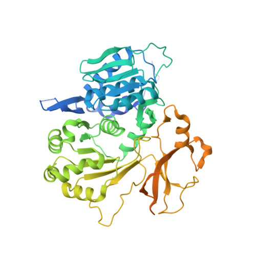
Macrolactam antibiotics such as incednine and cremimycin possess an aliphatic β-amino acid as a starter unit of their polyketide chain. In the biosynthesis of incednine and cremimycin, unique stand-alone adenylation enzymes IdnL1 and CmiS6 select and activate the proper aliphatic β-amino acid as a starter unit. In this study, we describe the enzymatic characterization and the structural basis of substrate specificity of IdnL1 and CmiS6. Functional analysis revealed that IdnL1 and CmiS6 recognize 3-aminobutanoic acid and 3-aminononanoic acid, respectively. We solved the X-ray crystal structures of IdnL1 and CmiS6 to understand the recognition mechanism of these aliphatic β-amino acids. These structures revealed that IdnL1 and CmiS6 share a common recognition motif that interacts with the β-amino group of the substrates. However, the hydrophobic side-chains of the substrates are accommodated differently in the two enzymes. IdnL1 has a bulky Leu220 located close to the terminal methyl group of 3-aminobutanoate of the trapped acyl-adenylate intermediate to construct a shallow substrate-binding pocket. In contrast, CmiS6 possesses Gly220 at the corresponding position to accommodate 3-aminononanoic acid. This structural observation was supported by a mutational study. Thus, the size of amino acid residue at the 220 position is critical for the selection of an aliphatic β-amino acid substrate in these adenylation enzymes. Proteins 2017; 85:1238-1247. © 2017 Wiley Periodicals, Inc.
- Department of Chemistry and Materials Science, Tokyo Institute of Technology, O-okayama, Meguro-ku, Tokyo, 152-8551, Japan.
Organizational Affiliation: