5JJD

crystal structure of the ceramide transfer protein PH and START domain complex


Experimental Data Snapshot

  • Method: X-RAY DIFFRACTION
  • Resolution: 2.40 Å
  • R-Value Free: 0.245 
  • R-Value Work: 0.201 
  • R-Value Observed: 0.203 

wwPDB Validation   3D Report Full Report


This is version 1.4 of the entry. See complete history


Literature

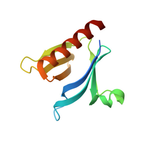
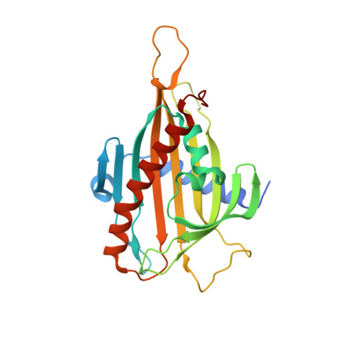
Interaction between the PH and START domains of ceramide transfer protein competes with phosphatidylinositol 4-phosphate binding by the PH domain.

Prashek, J.Bouyain, S.Fu, M.Li, Y.Berkes, D.Yao, X.

(2017) J Biol Chem 292: 14217-14228

  • DOI: https://doi.org/10.1074/jbc.M117.780007
  • Primary Citation of Related Structures:  
    5JJD

  • PubMed Abstract: 

    De novo synthesis of the sphingolipid sphingomyelin requires non-vesicular transport of ceramide from the endoplasmic reticulum to the Golgi by the multidomain protein ceramide transfer protein (CERT). CERT's N-terminal pleckstrin homology (PH) domain targets it to the Golgi by binding to phosphatidylinositol 4-phosphate (PtdIns(4)P) in the Golgi membrane, whereas its C-terminal StAR-related lipid transfer domain (START) carries out ceramide transfer. Hyperphosphorylation of a serine-rich motif immediately after the PH domain decreases both PtdIns(4)P binding and ceramide transfer by CERT. This down-regulation requires both the PH and START domains, suggesting a possible inhibitory interaction between the two domains. In this study we show that isolated PH and START domains interact with each other. The crystal structure of a PH-START complex revealed that the START domain binds to the PH domain at the same site for PtdIns(4)P-binding, suggesting that the START domain competes with PtdIns(4)P for association with the PH domain. We further report that mutations disrupting the PH-START interaction increase both PtdIns(4)P-binding affinity and ceramide transfer activity of a CERT-serine-rich phosphorylation mimic. We also found that these mutations increase the Golgi localization of CERT inside the cell, consistent with enhanced PtdIns(4)P binding of the mutant. Collectively, our structural, biochemical, and cellular investigations provide important structural insight into the regulation of CERT function and localization.


  • Organizational Affiliation

    From the Division of Molecular Biology and Biochemistry, School of Biological Sciences, University of Missouri-Kansas City, Kansas City, Missouri 64110.


Macromolecules
Find similar proteins by:  (by identity cutoff)  |  3D Structure
Entity ID: 1
MoleculeChains Sequence LengthOrganismDetailsImage
Collagen type IV alpha-3-binding protein106Homo sapiensMutation(s): 0 
Gene Names: COL4A3BPCERTSTARD11
UniProt & NIH Common Fund Data Resources
Find proteins for Q9Y5P4 (Homo sapiens)
Explore Q9Y5P4 
Go to UniProtKB:  Q9Y5P4
PHAROS:  Q9Y5P4
GTEx:  ENSG00000113163 
Entity Groups  
Sequence Clusters30% Identity50% Identity70% Identity90% Identity95% Identity100% Identity
UniProt GroupQ9Y5P4
Sequence Annotations
Expand
  • Reference Sequence
Find similar proteins by:  (by identity cutoff)  |  3D Structure
Entity ID: 2
MoleculeChains Sequence LengthOrganismDetailsImage
Collagen type IV alpha-3-binding protein246Homo sapiensMutation(s): 0 
Gene Names: COL4A3BPCERTSTARD11
UniProt & NIH Common Fund Data Resources
Find proteins for Q9Y5P4 (Homo sapiens)
Explore Q9Y5P4 
Go to UniProtKB:  Q9Y5P4
PHAROS:  Q9Y5P4
GTEx:  ENSG00000113163 
Entity Groups  
Sequence Clusters30% Identity50% Identity70% Identity90% Identity95% Identity100% Identity
UniProt GroupQ9Y5P4
Sequence Annotations
Expand
  • Reference Sequence
Experimental Data & Validation

Experimental Data

  • Method: X-RAY DIFFRACTION
  • Resolution: 2.40 Å
  • R-Value Free: 0.245 
  • R-Value Work: 0.201 
  • R-Value Observed: 0.203 
  • Space Group: P 21 21 21
Unit Cell:
Length ( Å )Angle ( ˚ )
a = 59.761α = 90
b = 60.914β = 90
c = 95.804γ = 90
Software Package:
Software NamePurpose
PHENIXrefinement
HKL-2000data reduction
HKL-2000data scaling
PHASERphasing

Structure Validation

View Full Validation Report


Entry History & Funding Information

Deposition Data


Funding OrganizationLocationGrant Number
National Institutes of Health/National Institute of General Medical Sciences (NIH/NIGMS)United States1 R15 GM113200-01

Revision History  (Full details and data files)

  • Version 1.0: 2017-07-05
    Type: Initial release
  • Version 1.1: 2017-09-13
    Changes: Author supporting evidence
  • Version 1.2: 2019-01-16
    Changes: Data collection, Database references
  • Version 1.3: 2019-12-25
    Changes: Author supporting evidence
  • Version 1.4: 2023-09-27
    Changes: Data collection, Database references, Refinement description