5JEX | pdb_00005jex

Crystal structure of type 2 PDF from Streptococcus agalactiae, crystallized in imidazole buffer


Experimental Data Snapshot

  • Method: X-RAY DIFFRACTION
  • Resolution: 2.00 Å
  • R-Value Free: 
    0.208 (Depositor), 0.209 (DCC) 
  • R-Value Work: 
    0.159 (Depositor), 0.168 (DCC) 
  • R-Value Observed: 
    0.161 (Depositor) 

Starting Model: experimental
View more details

wwPDB Validation 3D Report Full Report

Validation slider image for 5JEX

This is version 1.4 of the entry. See complete history

Literature

A unique peptide deformylase platform to rationally design and challenge novel active compounds.

Fieulaine, S.Alves de Sousa, R.Maigre, L.Hamiche, K.Alimi, M.Bolla, J.M.Taleb, A.Denis, A.Pages, J.M.Artaud, I.Meinnel, T.Giglione, C.

(2016) Sci Rep 6: 35429-35429

  • DOI: https://doi.org/10.1038/srep35429
  • Primary Citation Related Structures: 
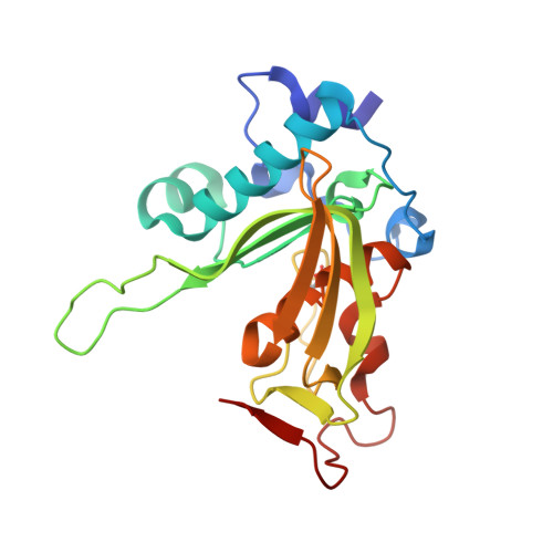
    5JEX, 5JEY, 5JEZ, 5JF0, 5JF1, 5JF2, 5JF3, 5JF4, 5JF5, 5JF6, 5JF7, 5JF8

  • PubMed Abstract: 

    Peptide deformylase (PDF) is considered an excellent target to develop antibiotics. We have performed an extensive characterization of a new PDF from the pathogen Streptococcus agalactiae, showing properties similar to other known PDFs. S. agalactiae PDF could be used as PDF prototype as it allowed to get complete sets of 3-dimensional, biophysical and kinetic data with virtually any inhibitor compound. Structure-activity relationship analysis with this single reference system allowed us to reveal distinct binding modes for different PDF inhibitors and the key role of a hydrogen bond in potentiating the interaction between ligand and target. We propose this protein as an irreplaceable tool, allowing easy and relevant fine comparisons between series, to design, challenge and validate novel series of inhibitors. As proof-of-concept, we report here the design and synthesis of effective specific bacterial PDF inhibitors of an oxadiazole series with potent antimicrobial activity against a multidrug resistant clinical isolate.


  • Organizational Affiliation
    • Institute for Integrative Biology of the Cell (I2BC), CEA, CNRS, Univ. Paris-Sud, Université Paris-Saclay, 91198 Gif-sur-Yvette cedex, France.

Macromolecule Content 

  • Total Structure Weight: 23.55 kDa 
  • Atom Count: 1,914 
  • Modeled Residue Count: 203 
  • Deposited Residue Count: 204 
  • Unique protein chains: 1

Macromolecules

Find similar proteins by:|  3D Structure
Entity ID: 1
MoleculeChains  Sequence LengthOrganismDetailsImage
Peptide deformylase204Streptococcus agalactiaeMutation(s): 1 
Gene Names: defgbs1883
EC: 3.5.1.88
UniProt
Find proteins for Q8E378 (Streptococcus agalactiae serotype III (strain NEM316))
Explore Q8E378 
Go to UniProtKB:  Q8E378
Entity Groups
Sequence Clusters30% Identity50% Identity70% Identity90% Identity95% Identity100% Identity
UniProt GroupQ8E378
Sequence Annotations
Expand
Reference Sequence

Small Molecules

Ligands 2 Unique
IDChains Name / Formula / InChI Key2D Diagram3D Interactions
IMD

Query on IMD


Download:Ideal Coordinates CCD File
B [auth A]IMIDAZOLE
C3 H5 N2
RAXXELZNTBOGNW-UHFFFAOYSA-O
ZN

Query on ZN


Download:Ideal Coordinates CCD File
C [auth A]
D [auth A]
E [auth A]
F [auth A]
G [auth A]
C [auth A],
D [auth A],
E [auth A],
F [auth A],
G [auth A],
H [auth A],
I [auth A],
J [auth A],
K [auth A]
ZINC ION
Zn
PTFCDOFLOPIGGS-UHFFFAOYSA-N
Modified Residues  1 Unique
IDChains TypeFormula2D DiagramParent
OCS
Query on OCS
A
L-PEPTIDE LINKINGC3 H7 N O5 SCYS

Experimental Data & Validation

Experimental Data

  • Method: X-RAY DIFFRACTION
  • Resolution: 2.00 Å
  • R-Value Free:  0.208 (Depositor), 0.209 (DCC) 
  • R-Value Work:  0.159 (Depositor), 0.168 (DCC) 
  • R-Value Observed: 0.161 (Depositor) 
Space Group: P 21 21 21
Unit Cell:
Length ( Å )Angle ( ˚ )
a = 41.32α = 90
b = 65.55β = 90
c = 88.71γ = 90
Software Package:
Software NamePurpose
REFMACrefinement
XDSdata reduction
XDSdata scaling
FRODOmodel building
PHASERphasing

Structure Validation

View Full Validation Report


Entry History 

& Funding Information

Deposition Data


Funding OrganizationLocationGrant Number
French National Research AgencyFranceANR6MIME 2006

Revision History  (Full details and data files)

  • Version 1.0: 2016-11-30
    Type: Initial release
  • Version 1.1: 2017-09-06
    Changes: Author supporting evidence
  • Version 1.2: 2018-04-25
    Changes: Data collection
  • Version 1.3: 2018-11-21
    Changes: Data collection
  • Version 1.4: 2024-01-10
    Changes: Data collection, Database references, Derived calculations, Refinement description