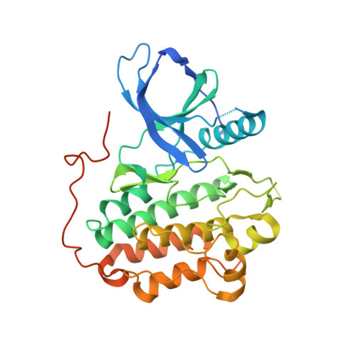
5JEB

Crystal structure of EGFR tyrosine kinase domain with novel inhibitor of active state of HER2


Experimental Data Snapshot

  • Method: X-RAY DIFFRACTION
  • Resolution: 3.30 Å
  • R-Value Free: 0.270 
  • R-Value Work: 0.242 
  • R-Value Observed: 0.244 

Starting Model: experimental
View more details

wwPDB Validation   3D Report Full Report


Ligand Structure Quality Assessment 


This is version 1.5 of the entry. See complete history


Literature

Overcoming resistance to HER2 inhibitors through state-specific kinase binding.

Novotny, C.J.Pollari, S.Park, J.H.Lemmon, M.A.Shen, W.Shokat, K.M.

(2016) Nat Chem Biol 12: 923-930

  • DOI: https://doi.org/10.1038/nchembio.2171
  • Primary Citation of Related Structures:  
    5JEB

  • PubMed Abstract: 

    The heterodimeric receptor tyrosine kinase complex formed by HER2 and HER3 can act as an oncogenic driver and is also responsible for rescuing a large number of cancers from a diverse set of targeted therapies. Inhibitors of these proteins, particularly HER2, have dramatically improved patient outcomes in the clinic, but recent studies have demonstrated that stimulating the heterodimeric complex, either via growth factors or by increasing the concentrations of HER2 and HER3 at the membrane, significantly diminishes the activity of the inhibitors. To identify an inhibitor of the active HER2-HER3 oncogenic complex, we developed a panel of Ba/F3 cell lines suitable for ultra-high-throughput screening. Medicinal chemistry on the hit scaffold resulted in a previously uncharacterized inhibitor that acts through preferential inhibition of the active state of HER2 and, as a result, is able to overcome cellular mechanisms of resistance such as growth factors or mutations that stabilize the active form of HER2.


  • Organizational Affiliation

    Howard Hughes Medical Institute, University of California San Francisco, San Francisco, California, USA.


Macromolecules
Find similar proteins by:  (by identity cutoff)  |  3D Structure
Entity ID: 1
MoleculeChains Sequence LengthOrganismDetailsImage
Epidermal growth factor receptor337Homo sapiensMutation(s): 1 
Gene Names: EGFRERBBERBB1HER1
EC: 2.7.10.1
UniProt & NIH Common Fund Data Resources
Find proteins for P00533 (Homo sapiens)
Explore P00533 
Go to UniProtKB:  P00533
PHAROS:  P00533
GTEx:  ENSG00000146648 
Entity Groups  
Sequence Clusters30% Identity50% Identity70% Identity90% Identity95% Identity100% Identity
UniProt GroupP00533
Sequence Annotations
Expand
  • Reference Sequence
Experimental Data & Validation

Experimental Data

  • Method: X-RAY DIFFRACTION
  • Resolution: 3.30 Å
  • R-Value Free: 0.270 
  • R-Value Work: 0.242 
  • R-Value Observed: 0.244 
  • Space Group: P 31 2 1
Unit Cell:
Length ( Å )Angle ( ˚ )
a = 69.435α = 90
b = 69.435β = 90
c = 191.52γ = 120
Software Package:
Software NamePurpose
PHENIXrefinement
PDB_EXTRACTdata extraction
HKL-2000data reduction
HKL-2000data scaling
PHASERphasing

Structure Validation

View Full Validation Report


Ligand Structure Quality Assessment 


Entry History & Funding Information

Deposition Data


Funding OrganizationLocationGrant Number
National Institutes of Health/National Institute of General Medical Sciences (NIH/NIGMS)United StatesR01-GM099891

Revision History  (Full details and data files)

  • Version 1.0: 2016-09-07
    Type: Initial release
  • Version 1.1: 2016-09-21
    Changes: Database references
  • Version 1.2: 2016-11-02
    Changes: Database references
  • Version 1.3: 2017-09-27
    Changes: Advisory, Author supporting evidence, Derived calculations
  • Version 1.4: 2019-12-25
    Changes: Author supporting evidence
  • Version 1.5: 2023-09-27
    Changes: Advisory, Data collection, Database references, Refinement description