5J9V

Ten minutes iron loaded Rana Catesbeiana H' ferritin variant E57A/E136A/D140A


Experimental Data Snapshot

  • Method: X-RAY DIFFRACTION
  • Resolution: 1.16 Å
  • R-Value Free: 0.138 
  • R-Value Work: 0.128 
  • R-Value Observed: 0.129 

wwPDB Validation   3D Report Full Report


This is version 1.5 of the entry. See complete history


Literature

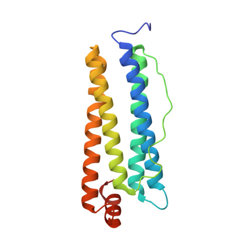
Ferroxidase Activity in Eukaryotic Ferritin is Controlled by Accessory-Iron-Binding Sites in the Catalytic Cavity.

Bernacchioni, C.Pozzi, C.Di Pisa, F.Mangani, S.Turano, P.

(2016) Chemistry 22: 16213-16219

  • DOI: https://doi.org/10.1002/chem.201602842
  • Primary Citation of Related Structures:  
    5J8S, 5J8W, 5J93, 5J9V, 5JAC

  • PubMed Abstract: 

    Ferritins are iron-storage nanocage proteins that catalyze the oxidation of Fe 2+ to Fe 3+ at ferroxidase sites. By a combination of structural and spectroscopic techniques, Asp140, together with previously identified Glu57 and Glu136, is demonstrated to be an essential residue to promote the iron oxidation at the ferroxidase site. However, the presence of these three carboxylate moieties in close proximity to the catalytic centers is not essential to achieve binding of the Fe 2+ substrate to the diferric ferroxidase sites with the same coordination geometries as in the wild-type cages.


  • Organizational Affiliation

    CERM and Department of Chemistry, University of Florence, Via Sacconi 6 Sesto Fiorentino, Firenze, 50019, Italy.


Macromolecules
Find similar proteins by:  (by identity cutoff)  |  3D Structure
Entity ID: 1
MoleculeChains Sequence LengthOrganismDetailsImage
Ferritin, middle subunit176Aquarana catesbeianaMutation(s): 3 
EC: 1.16.3.1
UniProt
Find proteins for P07798 (Aquarana catesbeiana)
Explore P07798 
Go to UniProtKB:  P07798
Entity Groups  
Sequence Clusters30% Identity50% Identity70% Identity90% Identity95% Identity100% Identity
UniProt GroupP07798
Sequence Annotations
Expand
  • Reference Sequence
Small Molecules
Ligands 3 Unique
IDChains Name / Formula / InChI Key2D Diagram3D Interactions
FE2
Query on FE2

Download Ideal Coordinates CCD File 
B [auth A]
C [auth A]
D [auth A]
E [auth A]
F [auth A]
B [auth A],
C [auth A],
D [auth A],
E [auth A],
F [auth A],
G [auth A],
H [auth A]
FE (II) ION
Fe
CWYNVVGOOAEACU-UHFFFAOYSA-N
CL
Query on CL

Download Ideal Coordinates CCD File 
AA [auth A]
BA [auth A]
CA [auth A]
DA [auth A]
EA [auth A]
AA [auth A],
BA [auth A],
CA [auth A],
DA [auth A],
EA [auth A],
O [auth A],
P [auth A],
Q [auth A],
R [auth A],
S [auth A],
T [auth A],
U [auth A],
V [auth A],
W [auth A],
X [auth A],
Y [auth A],
Z [auth A]
CHLORIDE ION
Cl
VEXZGXHMUGYJMC-UHFFFAOYSA-M
MG
Query on MG

Download Ideal Coordinates CCD File 
I [auth A]
J [auth A]
K [auth A]
L [auth A]
M [auth A]
I [auth A],
J [auth A],
K [auth A],
L [auth A],
M [auth A],
N [auth A]
MAGNESIUM ION
Mg
JLVVSXFLKOJNIY-UHFFFAOYSA-N
Experimental Data & Validation

Experimental Data

  • Method: X-RAY DIFFRACTION
  • Resolution: 1.16 Å
  • R-Value Free: 0.138 
  • R-Value Work: 0.128 
  • R-Value Observed: 0.129 
  • Space Group: F 4 3 2
Unit Cell:
Length ( Å )Angle ( ˚ )
a = 183.744α = 90
b = 183.744β = 90
c = 183.744γ = 90
Software Package:
Software NamePurpose
REFMACrefinement
XDSdata reduction
SCALAdata scaling
MOLREPphasing

Structure Validation

View Full Validation Report


Entry History 

Deposition Data

Revision History  (Full details and data files)

  • Version 1.0: 2016-10-05
    Type: Initial release
  • Version 1.1: 2016-11-02
    Changes: Database references
  • Version 1.2: 2017-11-29
    Changes: Database references
  • Version 1.3: 2018-01-31
    Changes: Database references
  • Version 1.4: 2019-02-20
    Changes: Data collection, Derived calculations
  • Version 1.5: 2024-01-10
    Changes: Data collection, Database references, Derived calculations, Refinement description