Penicillin V acylases from gram-negative bacteria degrade N-acylhomoserine lactones and attenuate virulence in Pseudomonas aeruginosa.
Sunder, A.V., Utari, P.D., Ramasamy, S., van Merkerk, R., Quax, W., Pundle, A.(2017) Appl Microbiol Biotechnol 101: 2383-2395
- PubMed: 27933456 Search on PubMed
- DOI: https://doi.org/10.1007/s00253-016-8031-5
- Primary Citation Related Structures:
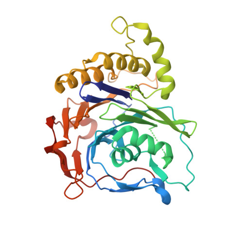
5J9R - PubMed Abstract:
Virulence pathways in gram-negative pathogenic bacteria are regulated by quorum sensing mechanisms, through the production and sensing of N-acylhomoserine lactone (AHL) signal molecules. Enzymatic degradation of AHLs leading to attenuation of virulence (quorum quenching) could pave the way for the development of new antibacterials. Penicillin V acylases (PVAs) belong to the Ntn hydrolase superfamily, together with AHL acylases. PVAs are exploited widely in the pharmaceutical industry, but their role in the natural physiology of their native microbes is not clearly understood. This report details the characterization of AHL degradation activity by homotetrameric PVAs from two gram-negative plant pathogenic bacteria, Pectobacterium atrosepticum (PaPVA) and Agrobacterium tumefaciens (AtPVA). Both the PVAs exhibited substrate specificity for degrading long-chain AHLs. Exogenous addition of these enzymes into Pseudomonas aeruginosa greatly diminished the production of elastase and pyocyanin and biofilm formation and increased the survival rate in an insect model of acute infection. Subtle structural differences in the PVA active site that regulate specificity for acyl chain length have been characterized, which could reflect the evolution of AHL-degrading acylases in relation to the environment of the bacteria that produce them and also provide strategies for enzyme engineering. The potential for using these enzymes as therapeutic agents in clinical applications and a few ideas about their possible significance in microbial physiology have also been discussed.
- Division of Biochemical Sciences, CSIR-National Chemical Laboratory, Dr. Homi Bhabha Road, Pune, 411008, India.
Organizational Affiliation: