5J4E

Crystal structures reveal signaling states of a short blue light photoreceptor protein PpSB1-LOV (Photoexcited state)


Experimental Data Snapshot

  • Method: X-RAY DIFFRACTION
  • Resolution: 2.67 Å
  • R-Value Free: 0.261 
  • R-Value Work: 0.186 
  • R-Value Observed: 0.190 

wwPDB Validation   3D Report Full Report


Ligand Structure Quality Assessment 


This is version 1.4 of the entry. See complete history


Literature

Signaling States of a Short Blue-Light Photoreceptor Protein PpSB1-LOV Revealed from Crystal Structures and Solution NMR Spectroscopy.

Rollen, K.Granzin, J.Panwalkar, V.Arinkin, V.Rani, R.Hartmann, R.Krauss, U.Jaeger, K.E.Willbold, D.Batra-Safferling, R.

(2016) J Mol Biol 428: 3721-3736

  • DOI: https://doi.org/10.1016/j.jmb.2016.05.027
  • Primary Citation of Related Structures:  
    5J3W, 5J4E

  • PubMed Abstract: 

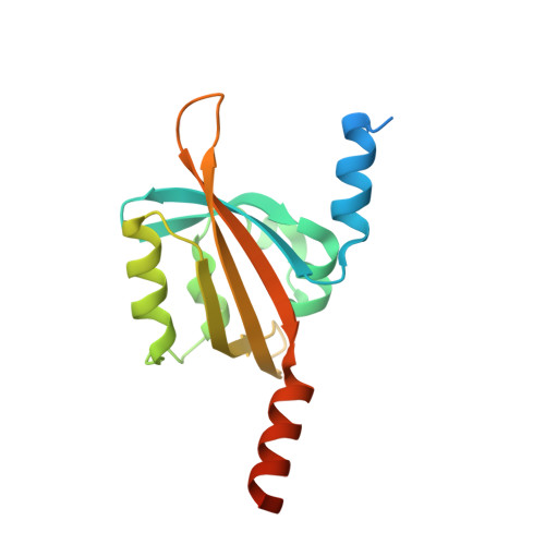
    Light-Oxygen-Voltage (LOV) domains represent the photo-responsive domains of various blue-light photoreceptor proteins and are widely distributed in plants, algae, fungi, and bacteria. Here, we report the dark-state crystal structure of PpSB1-LOV, a slow-reverting short LOV protein from Pseudomonas putida that is remarkably different from our previously published "fully light-adapted" structure [1]. A direct comparison of the two structures provides insight into the light-activated signaling mechanism. Major structural differences involve a~11Å movement of the C terminus in helix Jα, ~4Å movement of Hβ-Iβ loop, disruption of hydrogen bonds in the dimer interface, and a~29° rotation of chain-B relative to chain-A as compared to the light-state dimer. Both crystal structures and solution NMR data are suggestive of the key roles of a conserved glutamine Q116 and the N-cap region consisting of A'α-Aβ loop and the A'α helix in controlling the light-activated conformational changes. The activation mechanism proposed here for the PpSB1-LOV supports a rotary switch mechanism and provides insights into the signal propagation mechanism in naturally existing and artificial LOV-based, two-component systems and regulators.


  • Organizational Affiliation

    Institute of Complex Systems, ICS-6: Structural Biochemistry, Forschungszentrum Jülich, 52425 Jülich, Germany.


Macromolecules
Find similar proteins by:  (by identity cutoff)  |  3D Structure
Entity ID: 1
MoleculeChains Sequence LengthOrganismDetailsImage
Sensory box protein
A, B, C, D
162Pseudomonas putidaMutation(s): 0 
Gene Names: PP_4629
UniProt
Find proteins for Q88E39 (Pseudomonas putida (strain ATCC 47054 / DSM 6125 / CFBP 8728 / NCIMB 11950 / KT2440))
Explore Q88E39 
Go to UniProtKB:  Q88E39
Entity Groups  
Sequence Clusters30% Identity50% Identity70% Identity90% Identity95% Identity100% Identity
UniProt GroupQ88E39
Sequence Annotations
Expand
  • Reference Sequence
Experimental Data & Validation

Experimental Data

  • Method: X-RAY DIFFRACTION
  • Resolution: 2.67 Å
  • R-Value Free: 0.261 
  • R-Value Work: 0.186 
  • R-Value Observed: 0.190 
  • Space Group: C 1 2 1
Unit Cell:
Length ( Å )Angle ( ˚ )
a = 102.409α = 90
b = 71.08β = 91.02
c = 91.369γ = 90
Software Package:
Software NamePurpose
PHENIXrefinement
XDSdata reduction
Aimlessdata scaling
MOLREPphasing

Structure Validation

View Full Validation Report


Ligand Structure Quality Assessment 


Entry History & Funding Information

Deposition Data


Funding OrganizationLocationGrant Number
Bundesministerium fuer Bildung und ForschungGermanyFKZ-031A167B

Revision History  (Full details and data files)

  • Version 1.0: 2016-06-22
    Type: Initial release
  • Version 1.1: 2016-06-29
    Changes: Database references
  • Version 1.2: 2016-09-28
    Changes: Database references
  • Version 1.3: 2019-10-16
    Changes: Data collection
  • Version 1.4: 2024-01-10
    Changes: Data collection, Database references, Refinement description