5J2O

Crystal structure of the cyan fluorescence protein Cerulean S175G mutant


Experimental Data Snapshot

  • Method: X-RAY DIFFRACTION
  • Resolution: 1.50 Å
  • R-Value Free: 0.188 
  • R-Value Work: 0.169 

wwPDB Validation   3D Report Full Report


This is version 2.0 of the entry. See complete history


Literature

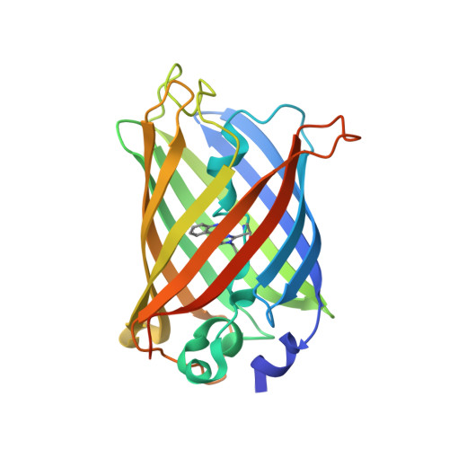
Crystal structure of the cyan fluorescent protein Cerulean-S175G

Park, S.-W.Kang, S.Yoon, T.-S.

(2016) Acta Crystallogr F Struct Biol Commun 72: 516-522

  • DOI: https://doi.org/10.1107/S2053230X16008311
  • Primary Citation of Related Structures:  
    5J2O

  • PubMed Abstract: 

    Enhanced cyan fluorescent protein (ECFP) was derived from Aequorea victoria green fluorescent protein (avGFP), notably with S65T/Y66W mutations. Its chromophore consists of a tripeptide comprised of Thr65, Trp66 and Gly67 (TWG) residues, while that of avGFP consists of a Ser65, Tyr66 and Gly67 (SYG) tripeptide. Cerulean and SCFP3A were derived from ECFP-S72A/H148D (a double mutation) with additional Y145A and S175G mutations, respectively, while Cerulean-S175G has both mutations (Y145A and S175G). The crystal structures of these ECFP variants at neutral pH were reported to adopt two distinct major conformations called ECFP and Cerulean. In this study, Cerulean-S175G was revealed to adopt only the Cerulean conformation, while Cerulean has been reported to adopt both the ECFP and the Cerulean conformations in its crystal structures. Sharing the same S175G mutation with SCFP3A, Cerulean-S175G showed a slightly increased quantum yield, like SCFP3A, but did not adopt the ECFP conformation adopted by SCFP3A. Detailed comparison of Cerulean-S175G and other ECFP variants revealed that the notable conformational changes in ECFP variants can be understood mainly in terms of the interaction between the Trp66 residue of the chromophore and residues 145-148 of β-strand 7.


  • Organizational Affiliation

    Bio-Analytical Science Major, Korea University of Science and Technology, KRIBB Campus, 125 Gwahak-ro, Yuseong-gu, Daejeon 34141, Republic of Korea.


Macromolecules
Find similar proteins by:  (by identity cutoff)  |  3D Structure
Entity ID: 1
MoleculeChains Sequence LengthOrganismDetailsImage
Green fluorescent protein243Aequorea victoriaMutation(s): 9 
Gene Names: GFP
UniProt
Find proteins for P42212 (Aequorea victoria)
Explore P42212 
Go to UniProtKB:  P42212
Entity Groups  
Sequence Clusters30% Identity50% Identity70% Identity90% Identity95% Identity100% Identity
UniProt GroupP42212
Sequence Annotations
Expand
  • Reference Sequence
Small Molecules
Modified Residues  1 Unique
IDChains TypeFormula2D DiagramParent
CRF
Query on CRF
A
L-PEPTIDE LINKINGC17 H18 N4 O4THR, TRP, GLY
Experimental Data & Validation

Experimental Data

  • Method: X-RAY DIFFRACTION
  • Resolution: 1.50 Å
  • R-Value Free: 0.188 
  • R-Value Work: 0.169 
  • Space Group: P 21 21 21
Unit Cell:
Length ( Å )Angle ( ˚ )
a = 51.137α = 90
b = 62.898β = 90
c = 70.106γ = 90
Software Package:
Software NamePurpose
PHENIXrefinement
PDB_EXTRACTdata extraction
Cootmodel building
HKL-2000data scaling
PHENIXphasing

Structure Validation

View Full Validation Report


Entry History & Funding Information

Deposition Data


Funding OrganizationLocationGrant Number
National Research Foundation of KoreaKorea, Republic OfNRF-2013M1A3A3A02042368

Revision History  (Full details and data files)

  • Version 1.0: 2016-04-13
    Type: Initial release
  • Version 1.1: 2016-07-20
    Changes: Database references
  • Version 1.2: 2023-11-08
    Changes: Data collection, Database references, Derived calculations, Refinement description
  • Version 2.0: 2023-11-15
    Changes: Atomic model, Data collection