5J0A | pdb_00005j0a

Crystal structure of PDZ-binding kinase


Experimental Data Snapshot

  • Method: X-RAY DIFFRACTION
  • Resolution: 2.74 Å
  • R-Value Free: 
    0.271 (Depositor), 0.273 (DCC) 
  • R-Value Work: 
    0.222 (Depositor), 0.226 (DCC) 
  • R-Value Observed: 
    0.222 (Depositor) 

wwPDB Validation 3D Report Full Report

Validation slider image for 5J0A

Ligand Structure Quality Assessment 


This is version 1.1 of the entry. See complete history

Literature

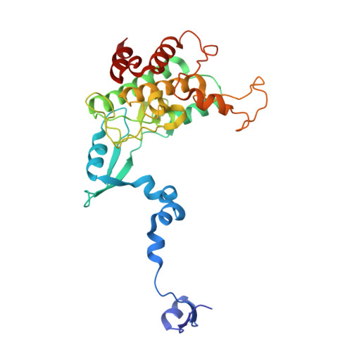
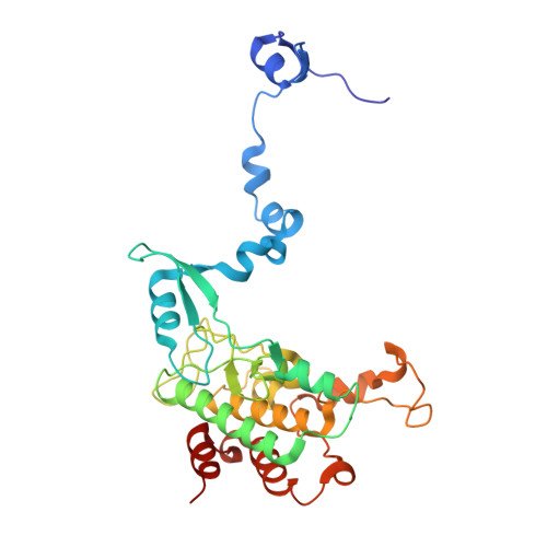
The crystal structure of an inactive dimer of PDZ-binding kinase

Dong, C.Tang, X.Xie, Y.Zou, Q.Yang, X.Zhou, H.

(2016) Biochem Biophys Res Commun 476: 586-593

  • DOI: https://doi.org/10.1016/j.bbrc.2016.05.166
  • Primary Citation Related Structures: 
    5J0A

  • PubMed Abstract: 

    The overexpression of PDZ-binding kinase/T-LAK cell-originated protein kinase (PBK/TOPK) has been associated with hematologic tumors, breast cancer and various other cancers. However, the three-dimensional structure of PBK has not been solved. In this study, we determined the crystal structure of human PBK, which has two phospho-mimicking mutations T9E and T198E. The structural data indicated that PBK may assemble into an inactive dimer in alkaline conditions. Analytical size-exclusion chromatography and analytical ultracentrifugation confirmed that PBK exists in a conformational transition between dimers and monomers at different pH conditions. Co-IP and kinase assays suggested that the active state of PBK is a monomer and does not form a dimer even under alkaline conditions. These results showed that the conformational transition of PBK is important for its kinase activity regulation. Collectively, our observations may provide a novel starting point for structure-based functional studies.


  • Organizational Affiliation
    • College of Life Sciences, Nankai University, 94 Weijin Road, Tianjin 300071, China.

Macromolecule Content 

  • Total Structure Weight: 69.25 kDa 
  • Atom Count: 4,975 
  • Modeled Residue Count: 602 
  • Deposited Residue Count: 604 
  • Unique protein chains: 2

Macromolecules

Find similar proteins by:|  3D Structure
Entity ID: 1
MoleculeChains  Sequence LengthOrganismDetailsImage
Lymphokine-activated killer T-cell-originated protein kinase300Homo sapiensMutation(s): 1 
Gene Names: PBKTOPK
EC: 2.7.12.2
UniProt & NIH Common Fund Data Resources
Find proteins for Q96KB5 (Homo sapiens)
Explore Q96KB5 
Go to UniProtKB:  Q96KB5
PHAROS:  Q96KB5
GTEx:  ENSG00000168078 
Entity Groups
Sequence Clusters30% Identity50% Identity70% Identity90% Identity95% Identity100% Identity
UniProt GroupQ96KB5
Sequence Annotations
Expand
Reference Sequence
Find similar proteins by:|  3D Structure
Entity ID: 2
MoleculeChains  Sequence LengthOrganismDetailsImage
Lymphokine-activated killer T-cell-originated protein kinase304Homo sapiensMutation(s): 1 
Gene Names: PBKTOPK
EC: 2.7.12.2
UniProt & NIH Common Fund Data Resources
Find proteins for Q96KB5 (Homo sapiens)
Explore Q96KB5 
Go to UniProtKB:  Q96KB5
PHAROS:  Q96KB5
GTEx:  ENSG00000168078 
Entity Groups
Sequence Clusters30% Identity50% Identity70% Identity90% Identity95% Identity100% Identity
UniProt GroupQ96KB5
Sequence Annotations
Expand
Reference Sequence

Small Molecules

Ligands 2 Unique
IDChains Name / Formula / InChI Key2D Diagram3D Interactions
HC4

Query on HC4


Download:Ideal Coordinates CCD File
H [auth A],
I [auth A],
J [auth A],
P [auth B]
4'-HYDROXYCINNAMIC ACID
C9 H8 O3
NGSWKAQJJWESNS-ZZXKWVIFSA-N
SO4

Query on SO4


Download:Ideal Coordinates CCD File
C [auth A]
D [auth A]
E [auth A]
F [auth A]
G [auth A]
C [auth A],
D [auth A],
E [auth A],
F [auth A],
G [auth A],
K [auth B],
L [auth B],
M [auth B],
N [auth B],
O [auth B]
SULFATE ION
O4 S
QAOWNCQODCNURD-UHFFFAOYSA-L

Experimental Data & Validation

Experimental Data

  • Method: X-RAY DIFFRACTION
  • Resolution: 2.74 Å
  • R-Value Free:  0.271 (Depositor), 0.273 (DCC) 
  • R-Value Work:  0.222 (Depositor), 0.226 (DCC) 
  • R-Value Observed: 0.222 (Depositor) 
Space Group: P 21 21 21
Unit Cell:
Length ( Å )Angle ( ˚ )
a = 75.558α = 90
b = 97.905β = 90
c = 162.835γ = 90
Software Package:
Software NamePurpose
CNSrefinement
HKL-2000data reduction
HKL-2000data scaling
PHENIXphasing

Structure Validation

View Full Validation Report


Ligand Structure Quality Assessment 


Entry History 

Deposition Data

Revision History  (Full details and data files)

  • Version 1.0: 2016-09-07
    Type: Initial release
  • Version 1.1: 2024-03-20
    Changes: Data collection, Database references, Derived calculations