5IWY | pdb_00005iwy

Crystal structure of 2-C-methyl-D-erythritol 2,4-cyclodiphosphate synthase from Bacillus subtitis complexed with CMP and Mg2+


Experimental Data Snapshot

  • Method: X-RAY DIFFRACTION
  • Resolution: 1.99 Å
  • R-Value Free: 
    0.219 (Depositor), 0.239 (DCC) 
  • R-Value Work: 
    0.174 (Depositor) 
  • R-Value Observed: 
    0.176 (Depositor) 

Starting Model: experimental
View more details

wwPDB Validation 3D Report Full Report

Validation slider image for 5IWY

Ligand Structure Quality Assessment 


This is version 1.2 of the entry. See complete history

Literature

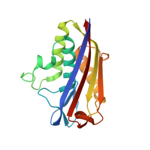
Crystal structure of IspF from Bacillus subtilis and absence of protein complex assembly among IspD/IspE/IspF enzymes in the MEP pathway.

Liu, Z.C.Jin, Y.Liu, W.F.Tao, Y.Wang, G.G.

(2018) Biosci Rep 

  • DOI: https://doi.org/10.1042/BSR20171370
  • Primary Citation Related Structures: 
    5IWX, 5IWY

  • PubMed Abstract: 

    2-C-Methyl-d-erythritol 2,4-cyclodiphosphate synthase (IspF) is a key enzyme in the 2-C-Methyl-d-erythritol-4-phosphate (MEP) pathway of isoprenoid biosynthesis. This enzyme catalyzes the 4-diphosphocytidyl-2-C-methyl-d-erythritol 2-phosphate (CDPME2P) to 2-C-methyl-d-erythritol 2,4-cyclodiphosphate (MEcDP) with concomitant release of cytidine 5'-diphospate (CMP). Bacillus subtilis is a potential host cell for the production of isoprenoids, but few studies are performed on the key enzymes of MEP pathway in B. subtilis In this work, the high-resolution crystal structures of IspF in native and complex with CMP from B. subtilis have been determined. Structural comparisons indicate that there is a looser packing of the subunits of IspF in B. subtilis , whereas the solvent accessible surface of its active pockets is smaller than that in Escherichia coli. Meanwhile, the protein-protein associations of 2-C-Methyl-d-erythritol-4-phosphatecytidyltransferase (IspD), CDPME kinase (IspE) and IspF from B. subtilis and E. coli , which catalyze three consecutive steps in the MEP pathway, are analyzed by native gel shift and size exclusion chromatography methods. The data here show that protein complex assembly is not detectable. These results will be useful for isoprenoid biosynthesis by metabolic engineering.


  • Organizational Affiliation
    • Key Laboratory of Environmental and Applied Microbiology, Chengdu Institute of Biology, Chinese Academy of Sciences, Chengdu 610041, China.

Macromolecule Content 

  • Total Structure Weight: 104.32 kDa 
  • Atom Count: 7,442 
  • Modeled Residue Count: 932 
  • Deposited Residue Count: 948 
  • Unique protein chains: 1

Macromolecules

Find similar proteins by:|  3D Structure
Entity ID: 1
MoleculeChains  Sequence LengthOrganismDetailsImage
2-C-methyl-D-erythritol 2,4-cyclodiphosphate synthase
A, B, C, D, E
A, B, C, D, E, F
158Bacillus subtilis subsp. subtilis str. 168Mutation(s): 0 
Gene Names: ispFyacNBSU00910
EC: 4.6.1.12
UniProt
Find proteins for Q06756 (Bacillus subtilis (strain 168))
Explore Q06756 
Go to UniProtKB:  Q06756
Entity Groups
Sequence Clusters30% Identity50% Identity70% Identity90% Identity95% Identity100% Identity
UniProt GroupQ06756
Sequence Annotations
Expand
Reference Sequence

Small Molecules

Ligands 2 Unique
IDChains Name / Formula / InChI Key2D Diagram3D Interactions
C5P

Query on C5P


Download:Ideal Coordinates CCD File
H [auth A],
I [auth B],
K [auth C],
O [auth F]
CYTIDINE-5'-MONOPHOSPHATE
C9 H14 N3 O8 P
IERHLVCPSMICTF-XVFCMESISA-N
MG

Query on MG


Download:Ideal Coordinates CCD File
G [auth A]
J [auth B]
L [auth C]
M [auth D]
N [auth E]
G [auth A],
J [auth B],
L [auth C],
M [auth D],
N [auth E],
P [auth F]
MAGNESIUM ION
Mg
JLVVSXFLKOJNIY-UHFFFAOYSA-N

Experimental Data & Validation

Experimental Data

  • Method: X-RAY DIFFRACTION
  • Resolution: 1.99 Å
  • R-Value Free:  0.219 (Depositor), 0.239 (DCC) 
  • R-Value Work:  0.174 (Depositor) 
  • R-Value Observed: 0.176 (Depositor) 
Space Group: P 1 21 1
Unit Cell:
Length ( Å )Angle ( ˚ )
a = 57.057α = 90
b = 89.46β = 103.8
c = 87.986γ = 90
Software Package:
Software NamePurpose
REFMACrefinement
HKL-2000data reduction
HKL-2000data scaling
PHASERphasing

Structure Validation

View Full Validation Report


Ligand Structure Quality Assessment 


Entry History 

Deposition Data

Revision History  (Full details and data files)

  • Version 1.0: 2017-03-29
    Type: Initial release
  • Version 1.1: 2018-02-14
    Changes: Database references
  • Version 1.2: 2023-11-08
    Changes: Data collection, Database references, Derived calculations, Refinement description