Effect of detergent binding on cytochrome P450 2B4 structure as analyzed by X-ray crystallography and deuterium-exchange mass spectrometry.
Shah, M.B., Jang, H.H., Wilderman, P.R., Lee, D., Li, S., Zhang, Q., Stout, C.D., Halpert, J.R.(2016) Biophys Chem 216: 1-8
- PubMed: 27280734 Search on PubMedSearch on PubMed Central
- DOI: https://doi.org/10.1016/j.bpc.2016.05.007
- Primary Citation Related Structures:
5IUT, 5IUZ - PubMed Abstract:
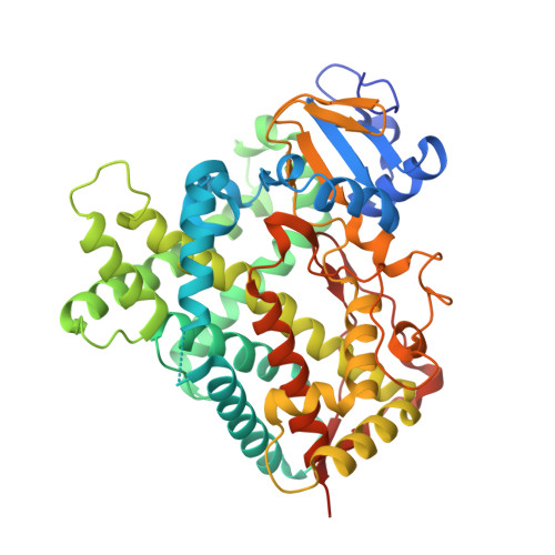
Multiple crystal structures of CYP2B4 have demonstrated the binding of the detergent 5-cyclohexyl-1-pentyl-β-D-maltoside (CYMAL-5) in a peripheral pocket located adjacent to the active site. To explore the consequences of detergent binding, X-ray crystal structures of the peripheral pocket mutant CYP2B4 F202W were solved in the presence of hexaethylene glycol monooctyl ether (C8E6) and CYMAL-5. The structure in the presence of CYMAL-5 illustrated a closed conformation indistinguishable from the previously solved wild-type. In contrast, the F202W structure in the presence of C8E6 revealed a detergent molecule that coordinated the heme-iron and extended to the protein surface through the substrate access channel 2f. Despite the overall structural similarity of these detergent complexes, remarkable differences were observed in the A, A', and H helices, the F-G cassette, the C-D and β4 loop region. Hydrogen-deuterium exchange mass spectrometry (DXMS) was employed to probe these differences and to test the effect of detergents in solution. The presence of either detergent increased the H/D exchange rate across the plastic regions, and the results obtained by DXMS in solution were consistent in general with the relevant structural snapshots. The study provides insight into effect of detergent binding and the interpretation of associated conformational dynamics of CYP2B4.
- School of Pharmacy, University of Connecticut, Storrs, CT 06269, United States. Electronic address: manish.shah@uconn.edu.
Organizational Affiliation: