5IUE | pdb_00005iue

Human leukocyte antigen F (HLA-F) presents peptides and regulates immunity through interactions with NK-cell receptors


Experimental Data Snapshot

  • Method: X-RAY DIFFRACTION
  • Resolution: 2.62 Å
  • R-Value Free: 
    0.241 (Depositor), 0.240 (DCC) 
  • R-Value Work: 
    0.197 (Depositor), 0.198 (DCC) 
  • R-Value Observed: 
    0.199 (Depositor) 

Starting Model: experimental
View more details

wwPDB Validation 3D Report Full Report

Validation slider image for 5IUE

Ligand Structure Quality Assessment 


This is version 1.6 of the entry. See complete history

Literature

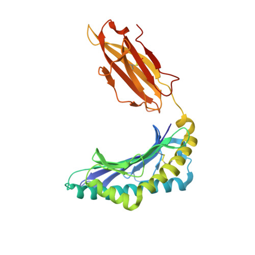
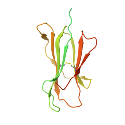
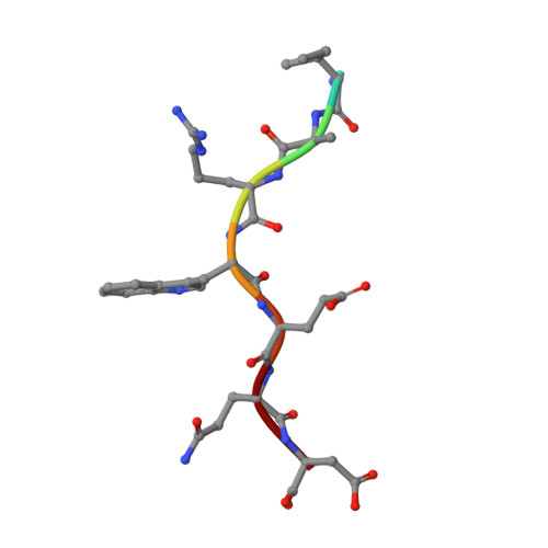
Human Leukocyte Antigen F Presents Peptides and Regulates Immunity through Interactions with NK Cell Receptors.

Dulberger, C.L.McMurtrey, C.P.Holzemer, A.Neu, K.E.Liu, V.Steinbach, A.M.Garcia-Beltran, W.F.Sulak, M.Jabri, B.Lynch, V.J.Altfeld, M.Hildebrand, W.H.Adams, E.J.

(2017) Immunity 46: 1018-1029.e7

  • DOI: https://doi.org/10.1016/j.immuni.2017.06.002
  • Primary Citation Related Structures: 
    5IUE, 5KNM

  • PubMed Abstract: 

    Evidence is mounting that the major histocompatibility complex (MHC) molecule HLA-F (human leukocyte antigen F) regulates the immune system in pregnancy, infection, and autoimmunity by signaling through NK cell receptors (NKRs). We present structural, biochemical, and evolutionary analyses demonstrating that HLA-F presents peptides of unconventional length dictated by a newly arisen mutation (R62W) that has produced an open-ended groove accommodating particularly long peptides. Compared to empty HLA-F open conformers (OCs), HLA-F tetramers bound with human-derived peptides differentially stained leukocytes, suggesting peptide-dependent engagement. Our in vitro studies confirm that NKRs differentiate between peptide-bound and peptide-free HLA-F. The complex structure of peptide-loaded β 2 m-HLA-F bound to the inhibitory LIR1 revealed similarities to high-affinity recognition of the viral MHC-I mimic UL18 and a docking strategy that relies on contacts with HLA-F as well as β 2 m, thus precluding binding to HLA-F OCs. These findings provide a biochemical framework to understand how HLA-F could regulate immunity via interactions with NKRs.


  • Organizational Affiliation
    • Department of Biochemistry and Molecular Biology, The University of Chicago, Chicago, IL 60637, USA.

Macromolecule Content 

  • Total Structure Weight: 215.83 kDa 
  • Atom Count: 12,729 
  • Modeled Residue Count: 1,525 
  • Deposited Residue Count: 1,896 
  • Unique protein chains: 3

Macromolecules

Find similar proteins by:|  3D Structure
Entity ID: 1
MoleculeChains  Sequence LengthOrganismDetailsImage
cDNA FLJ39643 fis, clone SMINT2004023, highly similar to HLA class I histocompatibility antigen, alphachain FA,
C [auth E],
E [auth G],
G [auth I]
284Homo sapiensMutation(s): 0 
UniProt & NIH Common Fund Data Resources
Find proteins for P30511 (Homo sapiens)
Explore P30511 
Go to UniProtKB:  P30511
PHAROS:  P30511
GTEx:  ENSG00000204642 
Entity Groups
Sequence Clusters30% Identity50% Identity70% Identity90% Identity95% Identity100% Identity
UniProt GroupP30511
Sequence Annotations
Expand
Reference Sequence
Find similar proteins by:|  3D Structure
Entity ID: 2
MoleculeChains  Sequence LengthOrganismDetailsImage
Beta-2-microglobulinB,
D [auth F],
F [auth H],
H [auth J]
182Homo sapiensMutation(s): 0 
Gene Names: B2MCDABP0092HDCMA22P
UniProt & NIH Common Fund Data Resources
Find proteins for P61769 (Homo sapiens)
Explore P61769 
Go to UniProtKB:  P61769
PHAROS:  P61769
GTEx:  ENSG00000166710 
Entity Groups
Sequence Clusters30% Identity50% Identity70% Identity90% Identity95% Identity100% Identity
UniProt GroupP61769
Sequence Annotations
Expand
Reference Sequence
Find similar proteins by:  Sequence   |   3D Structure  
Entity ID: 3
MoleculeChains  Sequence LengthOrganismDetailsImage
Peptide LEU-ILE-LEU-ARG-TRP-GLU-GLN-ASPI [auth K],
J [auth L],
K [auth M],
L [auth N]
8Trichoplusia niMutation(s): 0 

Small Molecules

Ligands 1 Unique
IDChains Name / Formula / InChI Key2D Diagram3D Interactions
NAG

Query on NAG


Download:Ideal Coordinates CCD File
M [auth I]2-acetamido-2-deoxy-beta-D-glucopyranose
C8 H15 N O6
OVRNDRQMDRJTHS-FMDGEEDCSA-N

Experimental Data & Validation

Experimental Data

  • Method: X-RAY DIFFRACTION
  • Resolution: 2.62 Å
  • R-Value Free:  0.241 (Depositor), 0.240 (DCC) 
  • R-Value Work:  0.197 (Depositor), 0.198 (DCC) 
  • R-Value Observed: 0.199 (Depositor) 
Space Group: P 21 21 21
Unit Cell:
Length ( Å )Angle ( ˚ )
a = 102.453α = 90
b = 115.324β = 90
c = 168.361γ = 90
Software Package:
Software NamePurpose
PHENIXrefinement
xia2data scaling
PHASERphasing

Structure Validation

View Full Validation Report


Ligand Structure Quality Assessment 


Entry History 

& Funding Information

Deposition Data


Funding OrganizationLocationGrant Number
National Institutes of Health/National Institute Of Allergy and Infectious Diseases (NIH/NIAID)United StatesR01AI073922

Revision History  (Full details and data files)

  • Version 1.0: 2017-06-14
    Type: Initial release
  • Version 1.1: 2017-07-05
    Changes: Database references
  • Version 1.2: 2017-09-27
    Changes: Author supporting evidence, Refinement description
  • Version 1.3: 2019-12-11
    Changes: Author supporting evidence
  • Version 1.4: 2020-07-29
    Type: Remediation
    Reason: Carbohydrate remediation
    Changes: Data collection, Derived calculations, Structure summary
  • Version 1.5: 2023-09-27
    Changes: Data collection, Database references, Refinement description, Structure summary
  • Version 1.6: 2024-11-20
    Changes: Structure summary