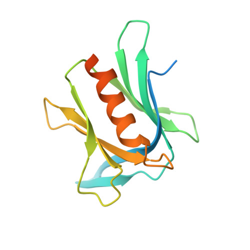
5IRS

crystal structure of the proteasomal Rpn13 PRU-domain


Experimental Data Snapshot

  • Method: X-RAY DIFFRACTION
  • Resolution: 1.80 Å
  • R-Value Free: 0.209 
  • R-Value Work: 0.167 
  • R-Value Observed: 0.171 

wwPDB Validation   3D Report Full Report


This is version 1.3 of the entry. See complete history


Literature

Structures of Rpn1 T1:Rad23 and hRpn13:hPLIC2 Reveal Distinct Binding Mechanisms between Substrate Receptors and Shuttle Factors of the Proteasome.

Chen, X.Randles, L.Shi, K.Tarasov, S.G.Aihara, H.Walters, K.J.

(2016) Structure 24: 1257-1270

  • DOI: https://doi.org/10.1016/j.str.2016.05.018
  • Primary Citation of Related Structures:  
    2NBU, 2NBV, 2NBW, 5IRS

  • PubMed Abstract: 

    Three receptors (Rpn1/S2/PSMD2, Rpn10/S5a, Rpn13/Adrm1) in the proteasome bind substrates by interacting with conjugated ubiquitin chains and/or shuttle factors (Rad23/HR23, Dsk2/PLIC/ubiquilin, Ddi1) that carry ubiquitinated substrates to proteasomes. We solved the structure of two such receptors with their preferred shuttle factor, namely hRpn13(Pru):hPLIC2(UBL) and scRpn1 T1:scRad23(UBL). We find that ubiquitin folds in Rad23 and Dsk2 are fine-tuned by residue substitutions to achieve high affinity for Rpn1 and Rpn13, respectively. A single substitution in hPLIC2 yields enhanced interactions with the Rpn13 ubiquitin contact surface and sterically blocks hRpn13 binding to its preferred ubiquitin chain type, K48-linked chains. Rpn1 T1 binds two ubiquitins in tandem and we find that Rad23 binds exclusively to the higher-affinity Helix28/Helix30 site. Rad23 contacts at Helix28/Helix30 are optimized compared to ubiquitin by multiple conservative amino acid substitutions. Thus, shuttle factors deliver substrates to proteasomes through fine-tuned ubiquitin-like surfaces.


  • Organizational Affiliation

    Protein Processing Section, Structural Biophysics Laboratory, Center for Cancer Research, National Cancer Institute, Frederick, MD 21702, USA.


Macromolecules
Find similar proteins by:  (by identity cutoff)  |  3D Structure
Entity ID: 1
MoleculeChains Sequence LengthOrganismDetailsImage
Proteasomal ubiquitin receptor ADRM1149Homo sapiensMutation(s): 0 
Gene Names: ADRM1GP110
UniProt & NIH Common Fund Data Resources
Find proteins for Q16186 (Homo sapiens)
Explore Q16186 
Go to UniProtKB:  Q16186
PHAROS:  Q16186
GTEx:  ENSG00000130706 
Entity Groups  
Sequence Clusters30% Identity50% Identity70% Identity90% Identity95% Identity100% Identity
UniProt GroupQ16186
Sequence Annotations
Expand
  • Reference Sequence
Small Molecules
Ligands 1 Unique
IDChains Name / Formula / InChI Key2D Diagram3D Interactions
DTT
Query on DTT

Download Ideal Coordinates CCD File 
B [auth A],
C [auth A]
2,3-DIHYDROXY-1,4-DITHIOBUTANE
C4 H10 O2 S2
VHJLVAABSRFDPM-IMJSIDKUSA-N
Experimental Data & Validation

Experimental Data

  • Method: X-RAY DIFFRACTION
  • Resolution: 1.80 Å
  • R-Value Free: 0.209 
  • R-Value Work: 0.167 
  • R-Value Observed: 0.171 
  • Space Group: P 21 21 21
Unit Cell:
Length ( Å )Angle ( ˚ )
a = 42.555α = 90
b = 56.476β = 90
c = 64.089γ = 90
Software Package:
Software NamePurpose
PHENIXrefinement
HKL-2000data reduction
HKL-2000data scaling
PHENIXphasing

Structure Validation

View Full Validation Report


Entry History 

Deposition Data

Revision History  (Full details and data files)

  • Version 1.0: 2016-07-20
    Type: Initial release
  • Version 1.1: 2016-08-03
    Changes: Data collection
  • Version 1.2: 2016-08-17
    Changes: Database references
  • Version 1.3: 2023-09-27
    Changes: Data collection, Database references, Derived calculations, Refinement description