5IMU | pdb_00005imu

A fragment of conserved hypothetical protein Rv3899c (residues 184-410) from Mycobacterium tuberculosis


Experimental Data Snapshot

  • Method: X-RAY DIFFRACTION
  • Resolution: 1.90 Å
  • R-Value Free: 
    0.190 (Depositor), 0.190 (DCC) 
  • R-Value Work: 
    0.160 (Depositor), 0.161 (DCC) 
  • R-Value Observed: 
    0.161 (Depositor) 

wwPDB Validation 3D Report Full Report

Validation slider image for 5IMU

This is version 1.1 of the entry. See complete history

Literature

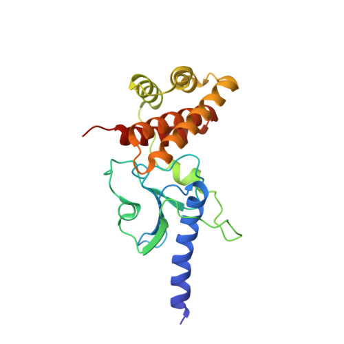
Crystal structure of Rv3899c(184-410), a hypothetical protein from Mycobacterium tuberculosis

Liu, Y.Y.Gao, Y.R.Li, D.F.Fleming, J.Li, H.L.Bi, L.J.

(2016) Acta Crystallogr F Struct Biol Commun 72: 642-645

  • DOI: https://doi.org/10.1107/S2053230X16010943
  • Primary Citation Related Structures: 
    5IMU

  • PubMed Abstract: 

    Rv3899c is a hypothetical protein from Mycobacterium tuberculosis which is conserved across mycobacteria. It is predicted to be secreted and has been found in culture filtrates. It has been proposed as a potential vaccine candidate; however, its biological function is unknown. Here, the global structure of Rv3899c(184-410), a fragment of Rv3899c, is reported. The structure resembles the shell of a sea snail, and its N- and C-termini form two relatively independent compact domains: an α/β/α sandwich folding domain and an α-helix bundle domain. There are no reported protein structures for any Rv3899c homologues; this structure provides the first structural glimpse of a new protein family consisting of Rv3899c and its homologues.


  • Organizational Affiliation
    • Shanghai Key Laboratory of New Drug Design, School of Pharmacy, East China University of Science and Technology, 130 Meilong Road, Shanghai 200237, People's Republic of China.

Macromolecule Content 

  • Total Structure Weight: 24.29 kDa 
  • Atom Count: 1,995 
  • Modeled Residue Count: 217 
  • Deposited Residue Count: 227 
  • Unique protein chains: 1

Macromolecules

Find similar proteins by:|  3D Structure
Entity ID: 1
MoleculeChains  Sequence LengthOrganismDetailsImage
Tat (Twin-arginine translocation) pathway signal sequence containing protein227Mycobacterium tuberculosis H37RvMutation(s): 0 
Gene Names: Rv3899cLH57_21230
UniProt
Find proteins for O05446 (Mycobacterium tuberculosis (strain ATCC 25618 / H37Rv))
Explore O05446 
Go to UniProtKB:  O05446
Entity Groups
Sequence Clusters30% Identity50% Identity70% Identity90% Identity95% Identity100% Identity
UniProt GroupO05446
Sequence Annotations
Expand
Reference Sequence

Small Molecules

Ligands 1 Unique
IDChains Name / Formula / InChI Key2D Diagram3D Interactions
K

Query on K


Download:Ideal Coordinates CCD File
B [auth A]POTASSIUM ION
K
NPYPAHLBTDXSSS-UHFFFAOYSA-N

Experimental Data & Validation

Experimental Data

  • Method: X-RAY DIFFRACTION
  • Resolution: 1.90 Å
  • R-Value Free:  0.190 (Depositor), 0.190 (DCC) 
  • R-Value Work:  0.160 (Depositor), 0.161 (DCC) 
  • R-Value Observed: 0.161 (Depositor) 
Space Group: P 21 21 21
Unit Cell:
Length ( Å )Angle ( ˚ )
a = 50.155α = 90
b = 54.973β = 90
c = 75.807γ = 90
Software Package:
Software NamePurpose
PHENIXrefinement
iMOSFLMdata reduction
SCALAdata scaling
PHENIXphasing

Structure Validation

View Full Validation Report


Entry History 

Deposition Data

Revision History  (Full details and data files)

  • Version 1.0: 2017-03-08
    Type: Initial release
  • Version 1.1: 2024-03-20
    Changes: Data collection, Database references