5IHE | pdb_00005ihe

D-family DNA polymerase - DP1 subunit (3'-5' proof-reading exonuclease)


Experimental Data Snapshot

  • Method: X-RAY DIFFRACTION
  • Resolution: 2.50 Å
  • R-Value Free: 
    0.225 (Depositor), 0.241 (DCC) 
  • R-Value Work: 
    0.197 (Depositor), 0.209 (DCC) 
  • R-Value Observed: 
    0.199 (Depositor) 

wwPDB Validation 3D Report Full Report

Validation slider image for 5IHE

Ligand Structure Quality Assessment 


This is version 1.2 of the entry. See complete history

Literature

Shared active site architecture between archaeal PolD and multi-subunit RNA polymerases revealed by X-ray crystallography.

Sauguet, L.Raia, P.Henneke, G.Delarue, M.

(2016) Nat Commun 7: 12227-12227

  • DOI: https://doi.org/10.1038/ncomms12227
  • Primary Citation Related Structures: 
    5IHE, 5IJL

  • PubMed Abstract: 

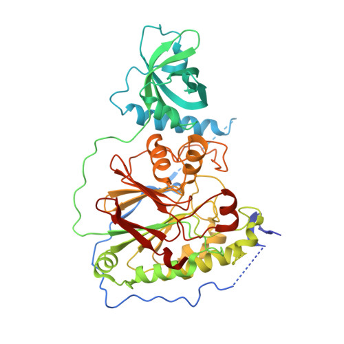
    Archaeal replicative DNA polymerase D (PolD) constitute an atypical class of DNA polymerases made of a proofreading exonuclease subunit (DP1) and a larger polymerase catalytic subunit (DP2), both with unknown structures. We have determined the crystal structures of Pyrococcus abyssi DP1 and DP2 at 2.5 and 2.2 Å resolution, respectively, revealing a catalytic core strikingly different from all other known DNA polymerases (DNAPs). Rather, the PolD DP2 catalytic core has the same 'double-psi β-barrel' architecture seen in the RNA polymerase (RNAP) superfamily, which includes multi-subunit transcriptases of all domains of life, homodimeric RNA-silencing pathway RNAPs and atypical viral RNAPs. This finding bridges together, in non-viral world, DNA transcription and DNA replication within the same protein superfamily. This study documents further the complex evolutionary history of the DNA replication apparatus in different domains of life and proposes a classification of all extant DNAPs.


  • Organizational Affiliation
    • Unit of Structural Dynamics of Macromolecules, Pasteur Institute and CNRS UMR 3528, 75015 Paris, France.

Macromolecule Content 

  • Total Structure Weight: 108.43 kDa 
  • Atom Count: 7,181 
  • Modeled Residue Count: 882 
  • Deposited Residue Count: 950 
  • Unique protein chains: 1

Macromolecules

Find similar proteins by:|  3D Structure
Entity ID: 1
MoleculeChains  Sequence LengthOrganismDetailsImage
DNA polymerase II small subunit
A, B
475Pyrococcus abyssi GE5Mutation(s): 0 
Gene Names: polBPYRAB01210PAB2266
EC: 2.7.7.7 (PDB Primary Data), 3.1.11.1 (UniProt)
UniProt
Find proteins for Q9V2F3 (Pyrococcus abyssi (strain GE5 / Orsay))
Explore Q9V2F3 
Go to UniProtKB:  Q9V2F3
Entity Groups
Sequence Clusters30% Identity50% Identity70% Identity90% Identity95% Identity100% Identity
UniProt GroupQ9V2F3
Sequence Annotations
Expand
Reference Sequence

Small Molecules

Ligands 6 Unique
IDChains Name / Formula / InChI Key2D Diagram3D Interactions
D5M

Query on D5M


Download:Ideal Coordinates CCD File
C [auth A],
I [auth B]
2'-DEOXYADENOSINE-5'-MONOPHOSPHATE
C10 H14 N5 O6 P
KHWCHTKSEGGWEX-RRKCRQDMSA-N
ZN

Query on ZN


Download:Ideal Coordinates CCD File
E [auth A],
K [auth B]
ZINC ION
Zn
PTFCDOFLOPIGGS-UHFFFAOYSA-N
EDO

Query on EDO


Download:Ideal Coordinates CCD File
N [auth B]1,2-ETHANEDIOL
C2 H6 O2
LYCAIKOWRPUZTN-UHFFFAOYSA-N
ACT

Query on ACT


Download:Ideal Coordinates CCD File
G [auth A],
M [auth B]
ACETATE ION
C2 H3 O2
QTBSBXVTEAMEQO-UHFFFAOYSA-M
FE

Query on FE


Download:Ideal Coordinates CCD File
D [auth A],
J [auth B]
FE (III) ION
Fe
VTLYFUHAOXGGBS-UHFFFAOYSA-N
CA

Query on CA


Download:Ideal Coordinates CCD File
F [auth A],
H [auth A],
L [auth B]
CALCIUM ION
Ca
BHPQYMZQTOCNFJ-UHFFFAOYSA-N

Experimental Data & Validation

Experimental Data

  • Method: X-RAY DIFFRACTION
  • Resolution: 2.50 Å
  • R-Value Free:  0.225 (Depositor), 0.241 (DCC) 
  • R-Value Work:  0.197 (Depositor), 0.209 (DCC) 
  • R-Value Observed: 0.199 (Depositor) 
Space Group: P 21 21 21
Unit Cell:
Length ( Å )Angle ( ˚ )
a = 85.969α = 90
b = 91.415β = 90
c = 145.209γ = 90
Software Package:
Software NamePurpose
BUSTERrefinement
XDSdata reduction
Aimlessdata scaling
AutoSolphasing

Structure Validation

View Full Validation Report


Ligand Structure Quality Assessment 


Entry History 

Deposition Data

Revision History  (Full details and data files)

  • Version 1.0: 2016-08-31
    Type: Initial release
  • Version 1.1: 2016-09-07
    Changes: Database references
  • Version 1.2: 2024-05-08
    Changes: Data collection, Database references, Derived calculations