5IE1 | pdb_00005ie1

Crystal structure of BACE1 in complex with 3-(2-amino-6-(o-tolyl)quinolin-3-yl)-N-(3,3-dimethylbutyl)propanamide


Experimental Data Snapshot

  • Method: X-RAY DIFFRACTION
  • Resolution: 2.30 Å
  • R-Value Free: 
    0.254 (Depositor), 0.257 (DCC) 
  • R-Value Work: 
    0.213 (Depositor), 0.216 (DCC) 
  • R-Value Observed: 
    0.216 (Depositor) 

Starting Model: other
View more details

wwPDB Validation 3D Report Full Report

Validation slider image for 5IE1

Ligand Structure Quality Assessment 


This is version 1.4 of the entry. See complete history

Literature

Fragment-Linking Approach Using (19)F NMR Spectroscopy To Obtain Highly Potent and Selective Inhibitors of beta-Secretase.

Jordan, J.B.Whittington, D.A.Bartberger, M.D.Sickmier, E.A.Chen, K.Cheng, Y.Judd, T.

(2016) J Med Chem 59: 3732-3749

  • DOI: https://doi.org/10.1021/acs.jmedchem.5b01917
  • Primary Citation Related Structures: 
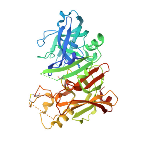
    5I3V, 5I3W, 5I3X, 5I3Y, 5IE1

  • PubMed Abstract: 

    Fragment-based drug discovery (FBDD) has become a widely used tool in small-molecule drug discovery efforts. One of the most commonly used biophysical methods in detecting weak binding of fragments is nuclear magnetic resonance (NMR) spectroscopy. In particular, FBDD performed with (19)F NMR-based methods has been shown to provide several advantages over (1)H NMR using traditional magnetization-transfer and/or two-dimensional methods. Here, we demonstrate the utility and power of (19)F-based fragment screening by detailing the identification of a second-site fragment through (19)F NMR screening that binds to a specific pocket of the aspartic acid protease, β-secretase (BACE-1). The identification of this second-site fragment allowed the undertaking of a fragment-linking approach, which ultimately yielded a molecule exhibiting a more than 360-fold increase in potency while maintaining reasonable ligand efficiency and gaining much improved selectivity over cathepsin-D (CatD). X-ray crystallographic studies of the molecules demonstrated that the linked fragments exhibited binding modes consistent with those predicted from the targeted screening approach, through-space NMR data, and molecular modeling.


  • Organizational Affiliation
    • Therapeutic Discovery, Amgen, Inc. One Amgen Center Drive, Thousand Oaks, California 91320, United States.

Macromolecule Content 

  • Total Structure Weight: 46.9 kDa 
  • Atom Count: 3,109 
  • Modeled Residue Count: 361 
  • Deposited Residue Count: 411 
  • Unique protein chains: 1

Macromolecules

Find similar proteins by:|  3D Structure
Entity ID: 1
MoleculeChains  Sequence LengthOrganismDetailsImage
Beta-secretase 1411Homo sapiensMutation(s): 0 
Gene Names: BACE1BACEKIAA1149
EC: 3.4.23.46
UniProt & NIH Common Fund Data Resources
Find proteins for P56817 (Homo sapiens)
Explore P56817 
Go to UniProtKB:  P56817
PHAROS:  P56817
GTEx:  ENSG00000186318 
Entity Groups
Sequence Clusters30% Identity50% Identity70% Identity90% Identity95% Identity100% Identity
UniProt GroupP56817
Sequence Annotations
Expand
Reference Sequence

Small Molecules

Ligands 3 Unique
IDChains Name / Formula / InChI Key2D Diagram3D Interactions
6BS

Query on 6BS


Download:Ideal Coordinates CCD File
H [auth A]3-[2-amino-6-(2-methylphenyl)quinolin-3-yl]-N-(3,3-dimethylbutyl)propanamide
C25 H31 N3 O
XKCSLMBNFSFIEK-UHFFFAOYSA-N
IOD

Query on IOD


Download:Ideal Coordinates CCD File
B [auth A],
C [auth A],
D [auth A],
E [auth A]
IODIDE ION
I
XMBWDFGMSWQBCA-UHFFFAOYSA-M
GOL

Query on GOL


Download:Ideal Coordinates CCD File
F [auth A],
G [auth A]
GLYCEROL
C3 H8 O3
PEDCQBHIVMGVHV-UHFFFAOYSA-N

Experimental Data & Validation

Experimental Data

  • Method: X-RAY DIFFRACTION
  • Resolution: 2.30 Å
  • R-Value Free:  0.254 (Depositor), 0.257 (DCC) 
  • R-Value Work:  0.213 (Depositor), 0.216 (DCC) 
  • R-Value Observed: 0.216 (Depositor) 
Space Group: P 61 2 2
Unit Cell:
Length ( Å )Angle ( ˚ )
a = 102.494α = 90
b = 102.494β = 90
c = 169.348γ = 120
Software Package:
Software NamePurpose
SCALEPACKdata scaling
PHENIXrefinement
PDB_EXTRACTdata extraction
HKL-2000data reduction
PHASERphasing

Structure Validation

View Full Validation Report


Ligand Structure Quality Assessment 


Entry History 

Deposition Data

Revision History  (Full details and data files)

  • Version 1.0: 2016-03-30
    Type: Initial release
  • Version 1.1: 2016-04-20
    Changes: Database references
  • Version 1.2: 2016-05-11
    Changes: Database references
  • Version 1.3: 2024-04-03
    Changes: Data collection, Database references, Derived calculations, Refinement description
  • Version 1.4: 2024-11-13
    Changes: Structure summary