


Crystal structure of an acyl-CoA dehydrogenase domain protein from Burkholderia phymatum bound to FAD
Edwards, T.E., Mayclin, S.J., Seattle Structural Genomics Center for Infectious Disease (SSGCID)To be published.
Experimental Data Snapshot
Starting Model: experimental
View more details
Entity ID: 1 | |||||
|---|---|---|---|---|---|
| Molecule | Chains | Sequence Length | Organism | Details | Image |
| Acyl-CoA dehydrogenase domain protein | 410 | Paraburkholderia phymatum STM815 | Mutation(s): 0 Gene Names: Bphy_4072 | 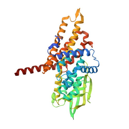 | |
UniProt | |||||
Entity Groups | |||||
| Sequence Clusters | 30% Identity50% Identity70% Identity90% Identity95% Identity100% Identity | ||||
| UniProt Group | B2JPK4 | ||||
Sequence AnnotationsExpand | |||||
Reference Sequence | |||||
| Ligands 2 Unique | |||||
|---|---|---|---|---|---|
| ID | Chains | Name / Formula / InChI Key | 2D Diagram | 3D Interactions | |
| FAD Download:Ideal Coordinates CCD File | E [auth A], I [auth B], M [auth C], Q [auth D] | FLAVIN-ADENINE DINUCLEOTIDE C27 H33 N9 O15 P2 VWWQXMAJTJZDQX-UYBVJOGSSA-N |  | ||
| EDO Download:Ideal Coordinates CCD File | F [auth A] G [auth A] H [auth A] J [auth B] K [auth B] | 1,2-ETHANEDIOL C2 H6 O2 LYCAIKOWRPUZTN-UHFFFAOYSA-N |  | ||
| Length ( Å ) | Angle ( ˚ ) |
|---|---|
| a = 84.18 | α = 90 |
| b = 108.42 | β = 90 |
| c = 189.44 | γ = 90 |
| Software Name | Purpose |
|---|---|
| PHENIX | refinement |
| XDS | data reduction |
| XSCALE | data scaling |
| PHASER | phasing |


