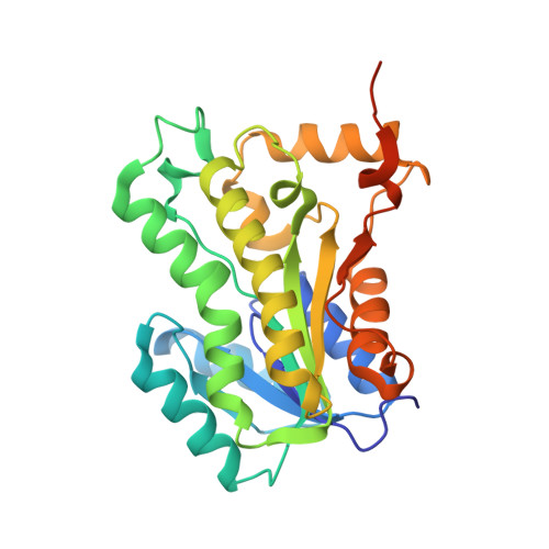
5I8Z

Crystal structure of B. pseudomallei FabI in complex with NAD and PT12


Experimental Data Snapshot

  • Method: X-RAY DIFFRACTION
  • Resolution: 1.62 Å
  • R-Value Free: 0.163 
  • R-Value Work: 0.141 
  • R-Value Observed: 0.142 

wwPDB Validation   3D Report Full Report


Ligand Structure Quality Assessment 


This is version 1.4 of the entry. See complete history


Literature

Rationalizing the Binding Kinetics for the Inhibition of the Burkholderia pseudomallei FabI1 Enoyl-ACP Reductase.

Neckles, C.Eltschkner, S.Cummings, J.E.Hirschbeck, M.Daryaee, F.Bommineni, G.R.Zhang, Z.Spagnuolo, L.Yu, W.Davoodi, S.Slayden, R.A.Kisker, C.Tonge, P.J.

(2017) Biochemistry 56: 1865-1878

  • DOI: https://doi.org/10.1021/acs.biochem.6b01048
  • Primary Citation of Related Structures:  
    5I7E, 5I7F, 5I7S, 5I7V, 5I8W, 5I8Z, 5I9L, 5I9M, 5I9N, 5IFL

  • PubMed Abstract: 

    There is growing awareness of the link between drug-target residence time and in vivo drug activity, and there are increasing efforts to determine the molecular factors that control the lifetime of a drug-target complex. Rational alterations in the drug-target residence time require knowledge of both the ground and transition states on the inhibition reaction coordinate, and we have determined the structure-kinetic relationship for 22 ethyl- or hexyl-substituted diphenyl ethers that are slow-binding inhibitors of bpFabI1, the enoyl-ACP reductase FabI1 from Burkholderia pseudomallei. Analysis of enzyme inhibition using a two-dimensional kinetic map demonstrates that the ethyl and hexyl diphenyl ethers fall into two distinct clusters. Modifications to the ethyl diphenyl ether B ring result in changes to both on and off rates, where residence times of up to ∼700 min (∼11 h) are achieved by either ground state stabilization (PT444) or transition state destabilization (slower on rate) (PT404). By contrast, modifications to the hexyl diphenyl ether B ring result in residence times of 300 min (∼5 h) through changes in only ground state stabilization (PT119). Structural analysis of nine enzyme:inhibitor complexes reveals that the variation in structure-kinetic relationships can be rationalized by structural rearrangements of bpFabI1 and subtle changes to the orientation of the inhibitor in the binding pocket. Finally, we demonstrate that three compounds with residence times on bpFabI1 from 118 min (∼2 h) to 670 min (∼11 h) have in vivo efficacy in an acute B. pseudomallei murine infection model using the virulent B. pseudomallei strain Bp400.


  • Organizational Affiliation

    Institute for Chemical Biology and Drug Discovery, Department of Chemistry, Stony Brook University , Stony Brook, New York 11794, United States.


Macromolecules
Find similar proteins by:  (by identity cutoff)  |  3D Structure
Entity ID: 1
MoleculeChains Sequence LengthOrganismDetailsImage
Enoyl-[acyl-carrier-protein] reductase [NADH]276Burkholderia pseudomalleiMutation(s): 0 
Gene Names: 
EC: 1.3.1.9
UniProt
Find proteins for A0A0H3HP34 (Burkholderia pseudomallei (strain 1026b))
Explore A0A0H3HP34 
Go to UniProtKB:  A0A0H3HP34
Entity Groups  
Sequence Clusters30% Identity50% Identity70% Identity90% Identity95% Identity100% Identity
UniProt GroupA0A0H3HP34
Sequence Annotations
Expand
  • Reference Sequence
Small Molecules
Ligands 2 Unique
IDChains Name / Formula / InChI Key2D Diagram3D Interactions
NAD
Query on NAD

Download Ideal Coordinates CCD File 
B [auth A]NICOTINAMIDE-ADENINE-DINUCLEOTIDE
C21 H27 N7 O14 P2
BAWFJGJZGIEFAR-NNYOXOHSSA-N
JA1
Query on JA1

Download Ideal Coordinates CCD File 
C [auth A]5-HEXYL-2-(4-NITROPHENOXY)PHENOL
C18 H21 N O4
NNNQXCYHIVPZRH-UHFFFAOYSA-N
Binding Affinity Annotations 
IDSourceBinding Affinity
JA1 Binding MOAD:  5I8Z Ki: 158 (nM) from 1 assay(s)
Experimental Data & Validation

Experimental Data

  • Method: X-RAY DIFFRACTION
  • Resolution: 1.62 Å
  • R-Value Free: 0.163 
  • R-Value Work: 0.141 
  • R-Value Observed: 0.142 
  • Space Group: I 2 2 2
Unit Cell:
Length ( Å )Angle ( ˚ )
a = 74.55α = 90
b = 75.91β = 90
c = 89.67γ = 90
Software Package:
Software NamePurpose
PHENIXrefinement
MOSFLMdata reduction
Aimlessdata scaling
PHASERphasing

Structure Validation

View Full Validation Report


Ligand Structure Quality Assessment 


Entry History & Funding Information

Deposition Data


Funding OrganizationLocationGrant Number
German Research FoundationGermanySFB 630

Revision History  (Full details and data files)

  • Version 1.0: 2017-02-22
    Type: Initial release
  • Version 1.1: 2017-03-08
    Changes: Database references
  • Version 1.2: 2017-04-19
    Changes: Database references
  • Version 1.3: 2017-09-06
    Changes: Author supporting evidence
  • Version 1.4: 2024-01-10
    Changes: Data collection, Database references, Refinement description