5I7Q | pdb_00005i7q

Crystal structure of Fkbp12-IF(SlpA), a chimeric protein of human Fkbp12 and the insert in flap domain of Ecoli SlpA


Experimental Data Snapshot

  • Method: X-RAY DIFFRACTION
  • Resolution: 1.90 Å
  • R-Value Free: 
    0.238 (Depositor), 0.238 (DCC) 
  • R-Value Work: 
    0.203 (Depositor), 0.206 (DCC) 
  • R-Value Observed: 
    0.205 (Depositor) 

Starting Model: experimental
View more details

wwPDB Validation 3D Report Full Report

Validation slider image for 5I7Q

This is version 1.2 of the entry. See complete history

Literature

Bacterial Chaperone Domain Insertions Convert Human FKBP12 into an Excellent Protein-Folding Catalyst-A Structural and Functional Analysis.

Zoldak, G.Knappe, T.A.Geitner, A.J.Scholz, C.Dobbek, H.Schmid, F.X.Jakob, R.P.

(2024) Molecules 29

  • DOI: https://doi.org/10.3390/molecules29071440
  • Primary Citation Related Structures: 
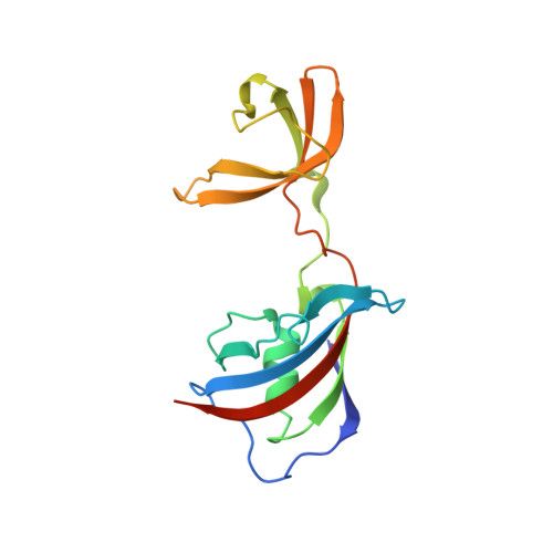
    5I7P, 5I7Q

  • PubMed Abstract: 

    Many folding enzymes use separate domains for the binding of substrate proteins and for the catalysis of slow folding reactions such as prolyl isomerization. FKBP12 is a small prolyl isomerase without a chaperone domain. Its folding activity is low, but it could be increased by inserting the chaperone domain from the homolog SlyD of E. coli near the prolyl isomerase active site. We inserted two other chaperone domains into human FKBP12: the chaperone domain of SlpA from E. coli , and the chaperone domain of SlyD from Thermococcus sp. Both stabilized FKBP12 and greatly increased its folding activity. The insertion of these chaperone domains had no influence on the FKBP12 and the chaperone domain structure, as revealed by two crystal structures of the chimeric proteins. The relative domain orientations differ in the two crystal structures, presumably representing snapshots of a more open and a more closed conformation. Together with crystal structures from SlyD-like proteins, they suggest a path for how substrate proteins might be transferred from the chaperone domain to the prolyl isomerase domain.


  • Organizational Affiliation
    • Center for Interdisciplinary Biosciences, Technology and Innovation Park, Pavol Jozef Šafárik University in Košice, 040 11 Kosice, Slovakia.

Macromolecule Content 

  • Total Structure Weight: 18.12 kDa 
  • Atom Count: 1,483 
  • Modeled Residue Count: 156 
  • Deposited Residue Count: 162 
  • Unique protein chains: 1

Macromolecules

Find similar proteins by:|  3D Structure
Entity ID: 1
MoleculeChains  Sequence LengthOrganismDetailsImage
Peptidyl-prolyl cis-trans isomerase FKBP1A,FKBP-type 16 kDa peptidyl-prolyl cis-trans isomerase,Peptidyl-prolyl cis-trans isomerase FKBP1A162Homo sapiensEscherichia coliMutation(s): 1 
Gene Names: FKBP1AFKBP1FKBP12fkpBslpAyaaDb0028JW0026
EC: 5.2.1.8
UniProt & NIH Common Fund Data Resources
Find proteins for P0AEM0 (Escherichia coli (strain K12))
Explore P0AEM0 
Go to UniProtKB:  P0AEM0
Find proteins for P62942 (Homo sapiens)
Explore P62942 
Go to UniProtKB:  P62942
PHAROS:  P62942
GTEx:  ENSG00000088832 
Entity Groups
Sequence Clusters30% Identity50% Identity70% Identity90% Identity95% Identity100% Identity
UniProt GroupsP62942P0AEM0
Sequence Annotations
Expand
Reference Sequence

Experimental Data & Validation

Experimental Data

  • Method: X-RAY DIFFRACTION
  • Resolution: 1.90 Å
  • R-Value Free:  0.238 (Depositor), 0.238 (DCC) 
  • R-Value Work:  0.203 (Depositor), 0.206 (DCC) 
  • R-Value Observed: 0.205 (Depositor) 
Space Group: P 21 21 2
Unit Cell:
Length ( Å )Angle ( ˚ )
a = 54.84α = 90
b = 63.61β = 90
c = 46.72γ = 90
Software Package:
Software NamePurpose
PHENIXrefinement
XDSdata reduction
XSCALEdata scaling
PHASERphasing

Structure Validation

View Full Validation Report


Entry History 

Deposition Data

Revision History  (Full details and data files)

  • Version 1.0: 2017-03-08
    Type: Initial release
  • Version 1.1: 2024-01-10
    Changes: Data collection, Database references, Refinement description
  • Version 1.2: 2026-04-22
    Changes: Database references, Structure summary