5I5U | pdb_00005i5u

X-RAY CRYSTAL STRUCTURE AT 2.40A RESOLUTION OF HUMAN MITOCHONDRIAL BRANCHED CHAIN AMINOTRANSFERASE (BCATM) COMPLEXED WITH A TETRAHYDRONAPHTHALENYL COMPOUND AND AN INTERNAL ALDIMINE LINKED PLP COFACTOR.


Experimental Data Snapshot

  • Method: X-RAY DIFFRACTION
  • Resolution: 2.40 Å
  • R-Value Free: 
    0.234 (Depositor), 0.237 (DCC) 
  • R-Value Work: 
    0.167 (Depositor), 0.176 (DCC) 
  • R-Value Observed: 
    0.171 (Depositor) 

Starting Model: other
View more details

wwPDB Validation 3D Report Full Report

Validation slider image for 5I5U

Ligand Structure Quality Assessment 


This is version 1.2 of the entry. See complete history

Literature

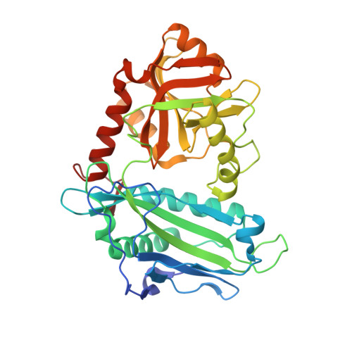
Structurally Diverse Mitochondrial Branched Chain Aminotransferase (BCATm) Leads with Varying Binding Modes Identified by Fragment Screening.

Borthwick, J.A.Ancellin, N.Bertrand, S.M.Bingham, R.P.Carter, P.S.Chung, C.W.Churcher, I.Dodic, N.Fournier, C.Francis, P.L.Hobbs, A.Jamieson, C.Pickett, S.D.Smith, S.E.Somers, D.O.Spitzfaden, C.Suckling, C.J.Young, R.J.

(2016) J Med Chem 59: 2452-2467

  • DOI: https://doi.org/10.1021/acs.jmedchem.5b01607
  • Primary Citation Related Structures: 
    5I5S, 5I5T, 5I5U, 5I5V, 5I5W, 5I5X, 5I5Y, 5I60

  • PubMed Abstract: 

    Inhibitors of mitochondrial branched chain aminotransferase (BCATm), identified using fragment screening, are described. This was carried out using a combination of STD-NMR, thermal melt (Tm), and biochemical assays to identify compounds that bound to BCATm, which were subsequently progressed to X-ray crystallography, where a number of exemplars showed significant diversity in their binding modes. The hits identified were supplemented by searching and screening of additional analogues, which enabled the gathering of further X-ray data where the original hits had not produced liganded structures. The fragment hits were optimized using structure-based design, with some transfer of information between series, which enabled the identification of ligand efficient lead molecules with micromolar levels of inhibition, cellular activity, and good solubility.


  • Organizational Affiliation
    • Medicines Research Centre, GlaxoSmithKline R&D , Gunnels Wood Road, Stevenage, Hertfordshire, SG1 2NY, U.K.

Macromolecule Content 

  • Total Structure Weight: 84.95 kDa 
  • Atom Count: 6,406 
  • Modeled Residue Count: 723 
  • Deposited Residue Count: 738 
  • Unique protein chains: 1

Macromolecules

Find similar proteins by:|  3D Structure
Entity ID: 1
MoleculeChains  Sequence LengthOrganismDetailsImage
Branched-chain-amino-acid aminotransferase, mitochondrial
A, B
369Homo sapiensMutation(s): 0 
Gene Names: BCAT2BCATMBCT2ECA40
EC: 2.6.1.42
UniProt & NIH Common Fund Data Resources
Find proteins for O15382 (Homo sapiens)
Explore O15382 
Go to UniProtKB:  O15382
PHAROS:  O15382
GTEx:  ENSG00000105552 
Entity Groups
Sequence Clusters30% Identity50% Identity70% Identity90% Identity95% Identity100% Identity
UniProt GroupO15382
Sequence Annotations
Expand
Reference Sequence

Small Molecules

Ligands 5 Unique
IDChains Name / Formula / InChI Key2D Diagram3D Interactions
PLP

Query on PLP


Download:Ideal Coordinates CCD File
C [auth A],
J [auth B]
PYRIDOXAL-5'-PHOSPHATE
C8 H10 N O6 P
NGVDGCNFYWLIFO-UHFFFAOYSA-N
67Y

Query on 67Y


Download:Ideal Coordinates CCD File
D [auth A],
K [auth B]
2-hydroxy-N-[(1R)-1,2,3,4-tetrahydronaphthalen-1-yl]acetamide
C12 H15 N O2
BBTOVZWCAMQWRJ-LLVKDONJSA-N
GOL

Query on GOL


Download:Ideal Coordinates CCD File
M [auth B]GLYCEROL
C3 H8 O3
PEDCQBHIVMGVHV-UHFFFAOYSA-N
EDO

Query on EDO


Download:Ideal Coordinates CCD File
F [auth A]
G [auth A]
H [auth A]
I [auth A]
N [auth B]
F [auth A],
G [auth A],
H [auth A],
I [auth A],
N [auth B],
O [auth B],
P [auth B]
1,2-ETHANEDIOL
C2 H6 O2
LYCAIKOWRPUZTN-UHFFFAOYSA-N
CL

Query on CL


Download:Ideal Coordinates CCD File
E [auth A],
L [auth B]
CHLORIDE ION
Cl
VEXZGXHMUGYJMC-UHFFFAOYSA-M

Experimental Data & Validation

Experimental Data

  • Method: X-RAY DIFFRACTION
  • Resolution: 2.40 Å
  • R-Value Free:  0.234 (Depositor), 0.237 (DCC) 
  • R-Value Work:  0.167 (Depositor), 0.176 (DCC) 
  • R-Value Observed: 0.171 (Depositor) 
Space Group: P 21 21 21
Unit Cell:
Length ( Å )Angle ( ˚ )
a = 69.074α = 90
b = 105.995β = 90
c = 107.462γ = 90
Software Package:
Software NamePurpose
SCALAdata scaling
REFMACrefinement
PDB_EXTRACTdata extraction
XDSdata reduction
REFMACphasing

Structure Validation

View Full Validation Report


Ligand Structure Quality Assessment 


Entry History 

Deposition Data

  • Released Date: 2016-03-23 
  • Deposition Author(s): Somers, D.O.

Revision History  (Full details and data files)

  • Version 1.0: 2016-03-23
    Type: Initial release
  • Version 1.1: 2016-04-06
    Changes: Database references
  • Version 1.2: 2024-05-01
    Changes: Data collection, Database references, Refinement description