5I4R | pdb_00005i4r

Contact-dependent inhibition system from Escherichia coli NC101 - ternary CdiA/CdiI/EF-Tu complex (trypsin-modified)


Experimental Data Snapshot

  • Method: X-RAY DIFFRACTION
  • Resolution: 3.30 Å
  • R-Value Free: 
    0.268 (Depositor), 0.234 (DCC) 
  • R-Value Work: 
    0.243 (Depositor) 
  • R-Value Observed: 
    0.244 (Depositor) 

Starting Models: experimental
View more details

wwPDB Validation 3D Report Full Report

Validation slider image for 5I4R

Ligand Structure Quality Assessment 


This is version 1.6 of the entry. See complete history

Literature

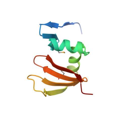
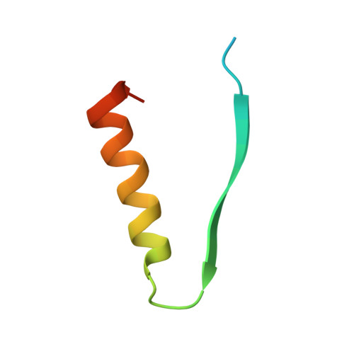
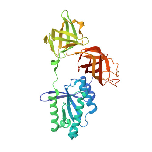
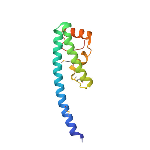
Structure of a novel antibacterial toxin that exploits elongation factor Tu to cleave specific transfer RNAs.

Michalska, K.Gucinski, G.C.Garza-Sanchez, F.Johnson, P.M.Stols, L.M.Eschenfeldt, W.H.Babnigg, G.Low, D.A.Goulding, C.W.Joachimiak, A.Hayes, C.S.

(2017) Nucleic Acids Res 45: 10306-10320

  • DOI: https://doi.org/10.1093/nar/gkx700
  • Primary Citation Related Structures: 
    5I4Q, 5I4R

  • PubMed Abstract: 

    Contact-dependent growth inhibition (CDI) is a mechanism of inter-cellular competition in which Gram-negative bacteria exchange polymorphic toxins using type V secretion systems. Here, we present structures of the CDI toxin from Escherichia coli NC101 in ternary complex with its cognate immunity protein and elongation factor Tu (EF-Tu). The toxin binds exclusively to domain 2 of EF-Tu, partially overlapping the site that interacts with the 3'-end of aminoacyl-tRNA (aa-tRNA). The toxin exerts a unique ribonuclease activity that cleaves the single-stranded 3'-end from tRNAs that contain guanine discriminator nucleotides. EF-Tu is required to support this tRNase activity in vitro, suggesting the toxin specifically cleaves substrate in the context of GTP·EF-Tu·aa-tRNA complexes. However, superimposition of the toxin domain onto previously solved GTP·EF-Tu·aa-tRNA structures reveals potential steric clashes with both aa-tRNA and the switch I region of EF-Tu. Further, the toxin induces conformational changes in EF-Tu, displacing a β-hairpin loop that forms a critical salt-bridge contact with the 3'-terminal adenylate of aa-tRNA. Together, these observations suggest that the toxin remodels GTP·EF-Tu·aa-tRNA complexes to free the 3'-end of aa-tRNA for entry into the nuclease active site.


  • Organizational Affiliation
    • Midwest Center for Structural Genomics, Biosciences Division, Argonne National Laboratory, Argonne, IL 60439, USA.

Macromolecule Content 

  • Total Structure Weight: 133.84 kDa 
  • Atom Count: 8,941 
  • Modeled Residue Count: 1,123 
  • Deposited Residue Count: 1,172 
  • Unique protein chains: 4

Macromolecules

Find similar proteins by:|  3D Structure
Entity ID: 1
MoleculeChains  Sequence LengthOrganismDetailsImage
Contact-dependent inhibitor A
A, E
92Escherichia coli NC101Mutation(s): 0 
EC: 3.1
UniProt
Find proteins for P0DSI1 (Escherichia coli (strain NC101))
Explore P0DSI1 
Go to UniProtKB:  P0DSI1
Entity Groups
Sequence Clusters30% Identity50% Identity70% Identity90% Identity95% Identity100% Identity
UniProt GroupP0DSI1
Sequence Annotations
Expand
Reference Sequence
Find similar proteins by:|  3D Structure
Entity ID: 2
MoleculeChains  Sequence LengthOrganismDetailsImage
Elongation factor TuB [auth C],
F [auth G]
45Escherichia coli BL21(DE3)Mutation(s): 0 
EC: 3.6.5.3
UniProt
Find proteins for P0CE48 (Escherichia coli (strain K12))
Explore P0CE48 
Go to UniProtKB:  P0CE48
Entity Groups
Sequence Clusters30% Identity50% Identity70% Identity90% Identity95% Identity100% Identity
UniProt GroupP0CE48
Sequence Annotations
Expand
Reference Sequence
Find similar proteins by:|  3D Structure
Entity ID: 3
MoleculeChains  Sequence LengthOrganismDetailsImage
Elongation factor TuC [auth D],
G [auth H]
335Escherichia coli BL21(DE3)Mutation(s): 0 
EC: 3.6.5.3
UniProt
Find proteins for P0CE47 (Escherichia coli (strain K12))
Explore P0CE47 
Go to UniProtKB:  P0CE47
Entity Groups
Sequence Clusters30% Identity50% Identity70% Identity90% Identity95% Identity100% Identity
UniProt GroupP0CE47
Sequence Annotations
Expand
Reference Sequence
Find similar proteins by:|  3D Structure
Entity ID: 4
MoleculeChains  Sequence LengthOrganismDetailsImage
Contact-dependent inhibitor ID [auth B],
H [auth F]
114Escherichia coli NC101Mutation(s): 0 
UniProt
Find proteins for P0DSM8 (Escherichia coli (strain NC101))
Explore P0DSM8 
Go to UniProtKB:  P0DSM8
Entity Groups
Sequence Clusters30% Identity50% Identity70% Identity90% Identity95% Identity100% Identity
UniProt GroupP0DSM8
Sequence Annotations
Expand
Reference Sequence

Small Molecules

Modified Residues  1 Unique
IDChains TypeFormula2D DiagramParent
MSE
Query on MSE
A, E
L-PEPTIDE LINKINGC5 H11 N O2 SeMET

Experimental Data & Validation

Experimental Data

  • Method: X-RAY DIFFRACTION
  • Resolution: 3.30 Å
  • R-Value Free:  0.268 (Depositor), 0.234 (DCC) 
  • R-Value Work:  0.243 (Depositor) 
  • R-Value Observed: 0.244 (Depositor) 
Space Group: P 1 21 1
Unit Cell:
Length ( Å )Angle ( ˚ )
a = 74.395α = 90
b = 128.354β = 109.58
c = 100.358γ = 90
Software Package:
Software NamePurpose
REFMACrefinement
HKL-3000data reduction
HKL-3000data scaling
PHASERphasing

Structure Validation

View Full Validation Report


Ligand Structure Quality Assessment 


Entry History 

& Funding Information

Deposition Data


Funding OrganizationLocationGrant Number
National Institutes of Health/National Institute of General Medical Sciences (NIH/NIGMS)United StatesGM094585
National Institutes of Health/National Institute of General Medical Sciences (NIH/NIGMS)United StatesGM102318

Revision History  (Full details and data files)

  • Version 1.0: 2017-06-28
    Type: Initial release
  • Version 1.1: 2017-09-20
    Changes: Author supporting evidence
  • Version 1.2: 2017-10-25
    Changes: Database references
  • Version 1.3: 2019-12-25
    Changes: Author supporting evidence
  • Version 1.4: 2023-09-27
    Changes: Data collection, Database references, Refinement description
  • Version 1.5: 2023-11-15
    Changes: Data collection
  • Version 1.6: 2024-10-30
    Changes: Structure summary