5I46 | pdb_00005i46

Factor VIIA in complex with the inhibitor (2R,15R)-2-[(1-aminoisoquinolin-6-yl)amino]-8-fluoro-7-hydroxy-4,15,17-trimethyl-13-oxa-4,11-diazatricyclo[14.2.2.1~6,10~]henicosa-1(18),6(21),7,9,16,19-hexaene-3,12-dione


Experimental Data Snapshot

  • Method: X-RAY DIFFRACTION
  • Resolution: 2.06 Å
  • R-Value Free: 
    0.239 (Depositor), 0.238 (DCC) 
  • R-Value Work: 
    0.217 (Depositor), 0.217 (DCC) 
  • R-Value Observed: 
    0.217 (Depositor) 

Starting Model: experimental
View more details

wwPDB Validation 3D Report Full Report

Validation slider image for 5I46

Ligand Structure Quality Assessment 


This is version 1.2 of the entry. See complete history

Literature

Atropisomer Control in Macrocyclic Factor VIIa Inhibitors.

Glunz, P.W.Mueller, L.Cheney, D.L.Ladziata, V.Zou, Y.Wurtz, N.R.Wei, A.Wong, P.C.Wexler, R.R.Priestley, E.S.

(2016) J Med Chem 59: 4007-4018

  • DOI: https://doi.org/10.1021/acs.jmedchem.6b00244
  • Primary Citation Related Structures: 
    5I46

  • PubMed Abstract: 

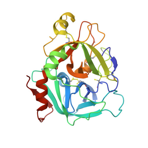
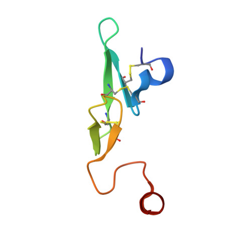
    Incorporation of a methyl group onto a macrocyclic FVIIa inhibitor improves potency 10-fold but is accompanied by atropisomerism due to restricted bond rotation in the macrocyclic structure, as demonstrated by NMR studies. We designed a conformational constraint favoring the desired atropisomer in which this methyl group interacts with the S2 pocket of FVIIa. A macrocyclic inhibitor incorporating this constraint was prepared and demonstrated by NMR to reside predominantly in the desired conformation. This modification improved potency 180-fold relative to the unsubstituted, racemic macrocycle and improved selectivity. An X-ray crystal structure of a closely related analogue in the FVIIa active site was obtained and matches the NMR and modeled conformations, confirming that this conformational constraint does indeed direct the methyl group into the S2 pocket as designed. The resulting rationally designed, conformationally stable template enables further optimization of these macrocyclic inhibitors.


  • Organizational Affiliation
    • Bristol-Myers Squibb Research & Development, 311 Pennington-Rocky Hill Road, Pennington, New Jersey 08534, United States.

Macromolecule Content 

  • Total Structure Weight: 35.2 kDa 
  • Atom Count: 2,690 
  • Modeled Residue Count: 309 
  • Deposited Residue Count: 309 
  • Unique protein chains: 2

Macromolecules

Find similar proteins by:|  3D Structure
Entity ID: 1
MoleculeChains  Sequence LengthOrganismDetailsImage
Coagulation factor VII (Heavy Chain)A [auth H]254Homo sapiensMutation(s): 0 
Gene Names: F7
EC: 3.4.21.21
UniProt & NIH Common Fund Data Resources
Find proteins for P08709 (Homo sapiens)
Explore P08709 
Go to UniProtKB:  P08709
PHAROS:  P08709
GTEx:  ENSG00000057593 
Entity Groups
Sequence Clusters30% Identity50% Identity70% Identity90% Identity95% Identity100% Identity
UniProt GroupP08709
Sequence Annotations
Expand
Reference Sequence
Find similar proteins by:|  3D Structure
Entity ID: 2
MoleculeChains  Sequence LengthOrganismDetailsImage
Coagulation factor VII (Light Chain)B [auth L]55Homo sapiensMutation(s): 0 
Gene Names: F7
EC: 3.4.21.21
UniProt & NIH Common Fund Data Resources
Find proteins for P08709 (Homo sapiens)
Explore P08709 
Go to UniProtKB:  P08709
PHAROS:  P08709
GTEx:  ENSG00000057593 
Entity Groups
Sequence Clusters30% Identity50% Identity70% Identity90% Identity95% Identity100% Identity
UniProt GroupP08709
Sequence Annotations
Expand
Reference Sequence

Small Molecules

Ligands 4 Unique
IDChains Name / Formula / InChI Key2D Diagram3D Interactions
67O

Query on 67O


Download:Ideal Coordinates CCD File
C [auth H](2R,15R)-2-[(1-aminoisoquinolin-6-yl)amino]-8-fluoro-7-hydroxy-4,15,17-trimethyl-13-oxa-4,11-diazatricyclo[14.2.2.1~6,10~]henicosa-1(18),6(21),7,9,16,19-hexaene-3,12-dione
C30 H30 F N5 O4
XABZFDVRWXGDRR-MRDQGFSESA-N
MES

Query on MES


Download:Ideal Coordinates CCD File
H
2-(N-MORPHOLINO)-ETHANESULFONIC ACID
C6 H13 N O4 S
SXGZJKUKBWWHRA-UHFFFAOYSA-N
SO4

Query on SO4


Download:Ideal Coordinates CCD File
E [auth H],
F [auth H],
G [auth H]
SULFATE ION
O4 S
QAOWNCQODCNURD-UHFFFAOYSA-L
CA

Query on CA


Download:Ideal Coordinates CCD File
D [auth H]CALCIUM ION
Ca
BHPQYMZQTOCNFJ-UHFFFAOYSA-N

Experimental Data & Validation

Experimental Data

  • Method: X-RAY DIFFRACTION
  • Resolution: 2.06 Å
  • R-Value Free:  0.239 (Depositor), 0.238 (DCC) 
  • R-Value Work:  0.217 (Depositor), 0.217 (DCC) 
  • R-Value Observed: 0.217 (Depositor) 
Space Group: P 41 21 2
Unit Cell:
Length ( Å )Angle ( ˚ )
a = 95.3α = 90
b = 95.3β = 90
c = 117.3γ = 90
Software Package:
Software NamePurpose
BUSTER-TNTrefinement
PDB_EXTRACTdata extraction
HKL-2000data reduction
HKL-2000data scaling
AMoREphasing

Structure Validation

View Full Validation Report


Ligand Structure Quality Assessment 


Entry History 

Deposition Data

  • Released Date: 2016-06-22 
  • Deposition Author(s): Wei, A.

Revision History  (Full details and data files)

  • Version 1.0: 2016-06-22
    Type: Initial release
  • Version 1.1: 2023-09-27
    Changes: Data collection, Database references, Derived calculations, Refinement description
  • Version 1.2: 2024-11-20
    Changes: Structure summary