5I30 | pdb_00005i30

Crystal structure of the human astrovirus 2 neutralizing monoclonal antibody PL-2 Fab fragment at 1.9 A resolution

  • Classification: IMMUNE SYSTEM
  • Organism(s): Mus musculus
  • Mutation(s): No 

  • Deposited: 2016-02-09 Released: 2016-04-13 
  • Deposition Author(s): Bogdanoff, W.A., DuBois, R.M.
  • Funding Organization(s): National Institutes of Health/National Institute Of Allergy and Infectious Diseases (NIH/NIAID)

Experimental Data Snapshot

  • Method: X-RAY DIFFRACTION
  • Resolution: 1.90 Å
  • R-Value Free: 
    0.221 (Depositor) 
  • R-Value Work: 
    0.180 (Depositor) 
  • R-Value Observed: 
    0.182 (Depositor) 

Starting Model: experimental
View more details

wwPDB Validation 3D Report Full Report

Validation slider image for 5I30

Ligand Structure Quality Assessment 


This is version 1.6 of the entry. See complete history

Literature

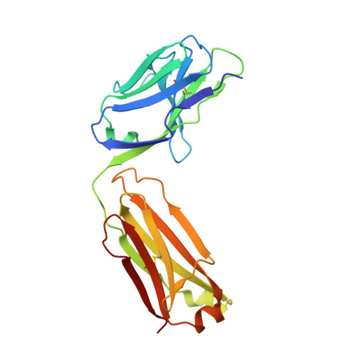
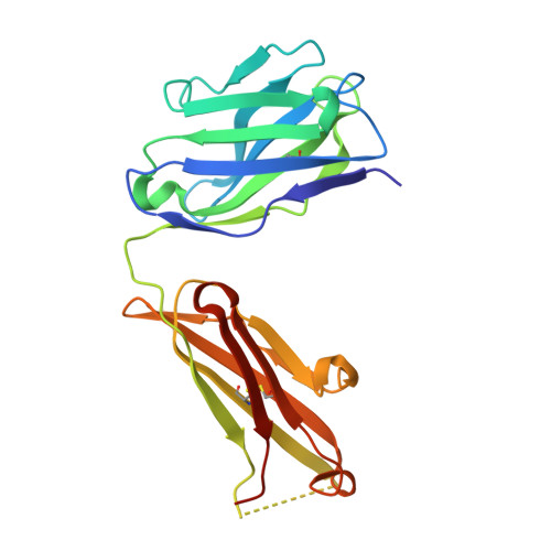
De Novo Sequencing and Resurrection of a Human Astrovirus-Neutralizing Antibody.

Bogdanoff, W.A.Morgenstern, D.Bern, M.Ueberheide, B.M.Sanchez-Fauquier, A.DuBois, R.M.

(2016) ACS Infect Dis 2: 313-321

  • DOI: https://doi.org/10.1021/acsinfecdis.6b00026
  • Primary Citation Related Structures: 
    5I30

  • PubMed Abstract: 

    Monoclonal antibody (mAb) therapeutics targeting cancer, autoimmune diseases, inflammatory diseases, and infectious diseases are growing exponentially. Although numerous panels of mAbs targeting infectious disease agents have been developed, their progression into clinically useful mAbs is often hindered by the lack of sequence information and/or loss of hybridoma cells that produce them. Here we combine the power of crystallography and mass spectrometry to determine the amino acid sequence and glycosylation modification of the Fab fragment of a potent human astrovirus-neutralizing mAb. We used this information to engineer a recombinant antibody single-chain variable fragment that has the same specificity as the parent monoclonal antibody to bind to the astrovirus capsid protein. This antibody can now potentially be developed as a therapeutic and diagnostic agent.


  • Organizational Affiliation
    • Department of Biomolecular Engineering, University of California Santa Cruz , 1156 High Street, Santa Cruz, California 95064, United States.

Macromolecule Content 

  • Total Structure Weight: 47.48 kDa 
  • Atom Count: 3,425 
  • Modeled Residue Count: 424 
  • Deposited Residue Count: 435 
  • Unique protein chains: 2

Macromolecules

Find similar proteins by:|  3D Structure
Entity ID: 1
MoleculeChains  Sequence LengthOrganismDetailsImage
Fab PL-2 light chainA [auth L]214Mus musculusMutation(s): 0 
Find similar proteins by:|  3D Structure
Entity ID: 2
MoleculeChains  Sequence LengthOrganismDetailsImage
Fab PL-2 heavy chainB [auth H]221Mus musculusMutation(s): 0 

Small Molecules

Ligands 1 Unique
IDChains Name / Formula / InChI Key2D Diagram3D Interactions
NAG

Query on NAG


Download:Ideal Coordinates CCD File
C [auth H]2-acetamido-2-deoxy-beta-D-glucopyranose
C8 H15 N O6
OVRNDRQMDRJTHS-FMDGEEDCSA-N

Experimental Data & Validation

Experimental Data

  • Method: X-RAY DIFFRACTION
  • Resolution: 1.90 Å
  • R-Value Free:  0.221 (Depositor) 
  • R-Value Work:  0.180 (Depositor) 
  • R-Value Observed: 0.182 (Depositor) 
Space Group: C 2 2 21
Unit Cell:
Length ( Å )Angle ( ˚ )
a = 72.22α = 90
b = 170.47β = 90
c = 78.27γ = 90
Software Package:
Software NamePurpose
PHENIXrefinement
MOSFLMdata reduction
SCALAdata scaling
Cootmodel building
PHASERphasing

Structure Validation

View Full Validation Report


Ligand Structure Quality Assessment 


Entry History 

& Funding Information

Deposition Data


Funding OrganizationLocationGrant Number
National Institutes of Health/National Institute Of Allergy and Infectious Diseases (NIH/NIAID)United StatesAI095369

Revision History  (Full details and data files)

  • Version 1.0: 2016-04-13
    Type: Initial release
  • Version 1.1: 2016-06-08
    Changes: Database references
  • Version 1.2: 2017-09-20
    Changes: Author supporting evidence, Derived calculations
  • Version 1.3: 2019-12-11
    Changes: Author supporting evidence, Database references
  • Version 1.4: 2020-07-29
    Type: Remediation
    Reason: Carbohydrate remediation
    Changes: Data collection, Derived calculations, Structure summary
  • Version 1.5: 2023-09-27
    Changes: Data collection, Database references, Refinement description, Structure summary
  • Version 1.6: 2024-10-23
    Changes: Structure summary