5I25

human recombinant coagulation FXI in complex with a peptide derived from human high molecular weight kininogen (HKP)


Experimental Data Snapshot

  • Method: X-RAY DIFFRACTION
  • Resolution: 2.85 Å
  • R-Value Free: 0.288 
  • R-Value Work: 0.226 
  • R-Value Observed: 0.232 

wwPDB Validation   3D Report Full Report


Ligand Structure Quality Assessment 


This is version 1.2 of the entry. See complete history


Literature

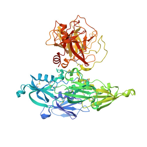
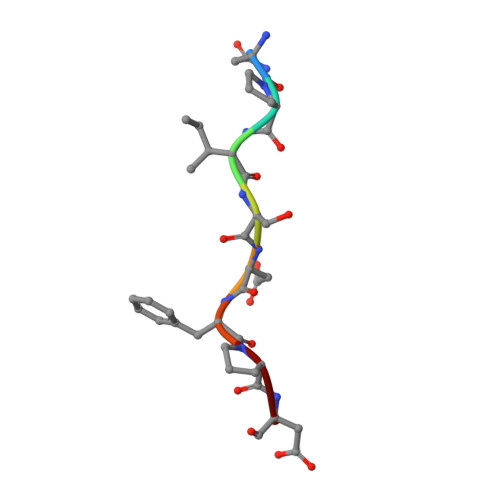
A novel DFP tripeptide motif interacts with the coagulation factor XI apple 2 domain.

Wong, S.S.stergaard, S.Hall, G.Li, C.Williams, P.M.Stennicke, H.Emsley, J.

(2016) Blood 127: 2915-2923

  • DOI: https://doi.org/10.1182/blood-2015-10-676122
  • Primary Citation of Related Structures:  
    5EOD, 5EOK, 5I25

  • PubMed Abstract: 
  • Factor XI (FXI) is the zymogen of FXIa, which cleaves FIX in the intrinsic pathway of coagulation. FXI is known to exist as a dimer and interacts with multiple proteins via its 4 apple domains in the "saucer section" of the enzyme; however, to date, no complex crystal structure has been described ...

    Factor XI (FXI) is the zymogen of FXIa, which cleaves FIX in the intrinsic pathway of coagulation. FXI is known to exist as a dimer and interacts with multiple proteins via its 4 apple domains in the "saucer section" of the enzyme; however, to date, no complex crystal structure has been described. To investigate protein interactions of FXI, a large random peptide library consisting of 10(6) to 10(7) peptides was screened for FXI binding, which identified a series of FXI binding motifs containing the signature Asp-Phe-Pro (DFP) tripeptide. Motifs containing this core tripeptide were found in diverse proteins, including the known ligand high-molecular-weight kininogen (HK), as well as the extracellular matrix proteins laminin and collagen V. To define the binding site on FXI, we determined the crystal structure of FXI in complex with the HK-derived peptide NPISDFPDT. This revealed the location of the DFP peptide bound to the FXI apple 2 domain, and central to the interaction, the DFP phenylalanine side-chain inserts into a major hydrophobic pocket in the apple 2 domain and the isoleucine occupies a flanking minor pocket. Two further structures of FXI in complex with the laminin-derived peptide EFPDFP and a DFP peptide from the random screen demonstrated binding in the same pocket, although in a slightly different conformation, thus revealing some flexibility in the molecular interactions of the FXI apple 2 domain.


    Organizational Affiliation

    Centre for Biomolecular Sciences, School of Pharmacy, University of Nottingham, Nottingham, United Kingdom; and.


Macromolecules
Find similar proteins by:  (by identity cutoff)  |  3D Structure
Entity ID: 1
MoleculeChainsSequence LengthOrganismDetailsImage
Coagulation factor XI607Homo sapiensMutation(s): 0 
Gene Names: F11
EC: 3.4.21.27
UniProt & NIH Common Fund Data Resources
Find proteins for P03951 (Homo sapiens)
Explore P03951 
Go to UniProtKB:  P03951
PHAROS:  P03951
Entity Groups  
Sequence Clusters30% Identity50% Identity70% Identity90% Identity95% Identity100% Identity
UniProt GroupP03951
Protein Feature View
Expand
  • Reference Sequence

Find similar proteins by:  Sequence   |   3D Structure  

Entity ID: 2
MoleculeChainsSequence LengthOrganismDetailsImage
ASN-PRO-ILE-SER-ASP-PHE-PRO-ASP8Homo sapiensMutation(s): 0 
Gene Names: KNG1BDKKNG
UniProt & NIH Common Fund Data Resources
Find proteins for P01042 (Homo sapiens)
Explore P01042 
Go to UniProtKB:  P01042
PHAROS:  P01042
Entity Groups  
UniProt GroupP01042
Protein Feature View
Expand
  • Reference Sequence
Small Molecules
Ligands 1 Unique
IDChainsName / Formula / InChI Key2D Diagram3D Interactions
NAG
Query on NAG

Download Ideal Coordinates CCD File 
C [auth A],
D [auth A],
E [auth A]
2-acetamido-2-deoxy-beta-D-glucopyranose
C8 H15 N O6
OVRNDRQMDRJTHS-FMDGEEDCSA-N
 Ligand Interaction
Experimental Data & Validation

Experimental Data

  • Method: X-RAY DIFFRACTION
  • Resolution: 2.85 Å
  • R-Value Free: 0.288 
  • R-Value Work: 0.226 
  • R-Value Observed: 0.232 
  • Space Group: P 43 21 2
Unit Cell:
Length ( Å )Angle ( ˚ )
a = 80.59α = 90
b = 80.59β = 90
c = 251.81γ = 90
Software Package:
Software NamePurpose
REFMACrefinement
XDSdata reduction
SCALAdata scaling
PHASERphasing

Structure Validation

View Full Validation Report


Ligand Structure Quality Assessment 


Entry History & Funding Information

Deposition Data


Funding OrganizationLocationGrant Number
British Heart FoundationUnited Kingdom--

Revision History  (Full details and data files)

  • Version 1.0: 2016-04-06
    Type: Initial release
  • Version 1.1: 2016-06-22
    Changes: Database references
  • Version 1.2: 2020-07-29
    Type: Remediation
    Reason: Carbohydrate remediation
    Changes: Data collection, Derived calculations, Structure summary