5I23 | pdb_00005i23

Crystal Structure of Agd31B, alpha-transglucosylase in Glycoside Hydrolase Family 31, in complex with Cyclophellitol Aziridine probe CF022


Experimental Data Snapshot

  • Method: X-RAY DIFFRACTION
  • Resolution: 1.95 Å
  • R-Value Free: 
    0.187 (Depositor), 0.194 (DCC) 
  • R-Value Work: 
    0.163 (Depositor), 0.172 (DCC) 
  • R-Value Observed: 
    0.164 (Depositor) 

Starting Model: experimental
View more details

wwPDB Validation 3D Report Full Report

Validation slider image for 5I23

Ligand Structure Quality Assessment 


This is version 1.3 of the entry. See complete history

Literature

Detection of Active Mammalian GH31 alpha-Glucosidases in Health and Disease Using In-Class, Broad-Spectrum Activity-Based Probes.

Jiang, J.Kuo, C.L.Wu, L.Franke, C.Kallemeijn, W.W.Florea, B.I.van Meel, E.van der Marel, G.A.Codee, J.D.Boot, R.G.Davies, G.J.Overkleeft, H.S.Aerts, J.M.

(2016) ACS Cent Sci 2: 351-358

  • DOI: https://doi.org/10.1021/acscentsci.6b00057
  • Primary Citation Related Structures: 
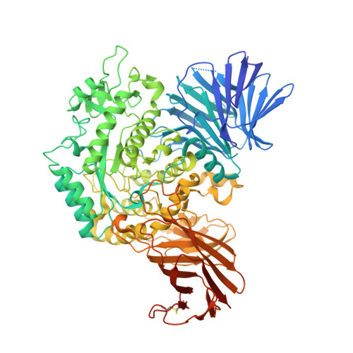
    5I23, 5I24

  • PubMed Abstract: 

    The development of small molecule activity-based probes (ABPs) is an evolving and powerful area of chemistry. There is a major need for synthetically accessible and specific ABPs to advance our understanding of enzymes in health and disease. α-Glucosidases are involved in diverse physiological processes including carbohydrate assimilation in the gastrointestinal tract, glycoprotein processing in the endoplasmic reticulum (ER), and intralysosomal glycogen catabolism. Inherited deficiency of the lysosomal acid α-glucosidase (GAA) causes the lysosomal glycogen storage disorder, Pompe disease. Here, we design a synthetic route for fluorescent and biotin-modified ABPs for in vitro and in situ monitoring of α-glucosidases. We show, through mass spectrometry, gel electrophoresis, and X-ray crystallography, that α-glucopyranose configured cyclophellitol aziridines label distinct retaining α-glucosidases including GAA and ER α-glucosidase II, and that this labeling can be tuned by pH. We illustrate a direct diagnostic application in Pompe disease patient cells, and discuss how the probes may be further exploited for diverse applications.


  • Organizational Affiliation
    • Department of Bio-organic Synthesis, Leiden Institute of Chemistry, Leiden University , Einsteinweg 55, 2333 CC Leiden, The Netherlands.

Macromolecule Content 

  • Total Structure Weight: 95.83 kDa 
  • Atom Count: 6,993 
  • Modeled Residue Count: 779 
  • Deposited Residue Count: 836 
  • Unique protein chains: 1

Macromolecules

Find similar proteins by:|  3D Structure
Entity ID: 1
MoleculeChains  Sequence LengthOrganismDetailsImage
Oligosaccharide 4-alpha-D-glucosyltransferase836Cellvibrio japonicus Ueda107Mutation(s): 0 
Gene Names: agd31BCJA_3248
EC: 2.4.1.161
UniProt
Find proteins for B3PEE6 (Cellvibrio japonicus (strain Ueda107))
Explore B3PEE6 
Go to UniProtKB:  B3PEE6
Entity Groups
Sequence Clusters30% Identity50% Identity70% Identity90% Identity95% Identity100% Identity
UniProt GroupB3PEE6
Sequence Annotations
Expand
Reference Sequence

Small Molecules

Ligands 4 Unique
IDChains Name / Formula / InChI Key2D Diagram3D Interactions
66V

Query on 66V


Download:Ideal Coordinates CCD File
K [auth A]1-(8-{[(1S,2R,3S,4S,5R,6R)-2,3,4,5-tetrahydroxy-6-(hydroxymethyl)cyclohexyl]amino}octyl)triaza-1,2-dien-2-ium
C15 H31 N4 O5
KPTZFIOZQLVZBJ-SAAWNECCSA-N
PG4

Query on PG4


Download:Ideal Coordinates CCD File
B [auth A]TETRAETHYLENE GLYCOL
C8 H18 O5
UWHCKJMYHZGTIT-UHFFFAOYSA-N
SO4

Query on SO4


Download:Ideal Coordinates CCD File
C [auth A]
D [auth A]
E [auth A]
F [auth A]
G [auth A]
C [auth A],
D [auth A],
E [auth A],
F [auth A],
G [auth A],
H [auth A]
SULFATE ION
O4 S
QAOWNCQODCNURD-UHFFFAOYSA-L
EDO

Query on EDO


Download:Ideal Coordinates CCD File
I [auth A],
J [auth A]
1,2-ETHANEDIOL
C2 H6 O2
LYCAIKOWRPUZTN-UHFFFAOYSA-N

Experimental Data & Validation

Experimental Data

  • Method: X-RAY DIFFRACTION
  • Resolution: 1.95 Å
  • R-Value Free:  0.187 (Depositor), 0.194 (DCC) 
  • R-Value Work:  0.163 (Depositor), 0.172 (DCC) 
  • R-Value Observed: 0.164 (Depositor) 
Space Group: P 6 2 2
Unit Cell:
Length ( Å )Angle ( ˚ )
a = 198α = 90
b = 198β = 90
c = 103γ = 120
Software Package:
Software NamePurpose
REFMACrefinement
xia2data reduction
xia2data scaling
MOLREPphasing

Structure Validation

View Full Validation Report


Ligand Structure Quality Assessment 


Entry History 

& Funding Information

Deposition Data


Funding OrganizationLocationGrant Number
European Research CouncilUnited KingdomERC-2012-AdG-322942

Revision History  (Full details and data files)

  • Version 1.0: 2016-05-04
    Type: Initial release
  • Version 1.1: 2016-06-22
    Changes: Database references
  • Version 1.2: 2024-01-10
    Changes: Data collection, Database references, Refinement description
  • Version 1.3: 2024-11-20
    Changes: Structure summary