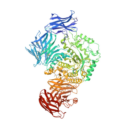
5I0D

Cycloalternan-forming enzyme from Listeria monocytogenes in complex with cycloalternan


Experimental Data Snapshot

  • Method: X-RAY DIFFRACTION
  • Resolution: 1.77 Å
  • R-Value Free: 0.171 
  • R-Value Work: 0.144 
  • R-Value Observed: 0.145 

Starting Model: experimental
View more details

wwPDB Validation   3D Report Full Report


Ligand Structure Quality Assessment 


This is version 2.3 of the entry. See complete history


Literature

Transferase Versus Hydrolase: The Role of Conformational Flexibility in Reaction Specificity.

Light, S.H.Cahoon, L.A.Mahasenan, K.V.Lee, M.Boggess, B.Halavaty, A.S.Mobashery, S.Freitag, N.E.Anderson, W.F.

(2017) Structure 25: 295-304

  • DOI: https://doi.org/10.1016/j.str.2016.12.007
  • Primary Citation of Related Structures:  
    5HOP, 5HPO, 5HXM, 5I0D, 5I0E, 5I0F, 5I0G

  • PubMed Abstract: 

    Active in the aqueous cellular environment where a massive excess of water is perpetually present, enzymes that catalyze the transfer of an electrophile to a non-water nucleophile (transferases) require specific strategies to inhibit mechanistically related hydrolysis reactions. To identify principles that confer transferase versus hydrolase reaction specificity, we exploited two enzymes that use highly similar catalytic apparatuses to catalyze the transglycosylation (a transferase reaction) or hydrolysis of α-1,3-glucan linkages in the cyclic tetrasaccharide cycloalternan (CA). We show that substrate binding to non-catalytic domains and a conformationally stable active site promote CA transglycosylation, whereas a distinct pattern of active site conformational change is associated with CA hydrolysis. These findings defy the classic view of induced-fit conformational change and illustrate a mechanism by which a stable hydrophobic binding site can favor transferase activity and disfavor hydrolysis. Application of these principles could facilitate the rational reengineering of transferases with desired catalytic properties.


  • Organizational Affiliation

    Department of Biochemistry and Molecular Genetics, Center for Structural Genomics of Infectious Diseases, Feinberg School of Medicine, Northwestern University, Chicago, IL 60611, USA.


Macromolecules
Find similar proteins by:  (by identity cutoff)  |  3D Structure
Entity ID: 1
MoleculeChains Sequence LengthOrganismDetailsImage
Lmo2446 protein
A, B
1,063Listeria monocytogenes EGD-eMutation(s): 0 
Gene Names: lmo2446
UniProt
Find proteins for Q8Y4J2 (Listeria monocytogenes serovar 1/2a (strain ATCC BAA-679 / EGD-e))
Explore Q8Y4J2 
Go to UniProtKB:  Q8Y4J2
Entity Groups  
Sequence Clusters30% Identity50% Identity70% Identity90% Identity95% Identity100% Identity
UniProt GroupQ8Y4J2
Sequence Annotations
Expand
  • Reference Sequence
Oligosaccharides

Help

Entity ID: 2
MoleculeChains Length2D Diagram Glycosylation3D Interactions
alpha-D-glucopyranose-(1-6)-alpha-D-glucopyranose
C, E, I, J
2N/A
Glycosylation Resources
GlyTouCan:  G69864PN
GlyCosmos:  G69864PN
Entity ID: 3
MoleculeChains Length2D Diagram Glycosylation3D Interactions
Cyclic alpha-D-glucopyranose-(1-3)-alpha-D-glucopyranose-(1-6)-alpha-D-glucopyranose-(1-3)-alpha-D-glucopyranose
D, F, K, L, M
4N/A
Glycosylation Resources
GlyTouCan:  G67006KT
GlyCosmos:  G67006KT
Entity ID: 4
MoleculeChains Length2D Diagram Glycosylation3D Interactions
alpha-D-glucopyranose-(1-3)-alpha-D-glucopyranose-(1-6)-alpha-D-glucopyranose
G
3N/A
Glycosylation Resources
GlyTouCan:  G92776DG
GlyCosmos:  G92776DG
GlyGen:  G92776DG
Entity ID: 5
MoleculeChains Length2D Diagram Glycosylation3D Interactions
Cyclic alpha-D-glucopyranose-(1-6)-alpha-D-glucopyranose-(1-3)-alpha-D-glucopyranose-(1-6)-alpha-D-glucopyranose
H
4N/A
Glycosylation Resources
GlyTouCan:  G32655OG
GlyCosmos:  G32655OG
Small Molecules
Ligands 4 Unique
IDChains Name / Formula / InChI Key2D Diagram3D Interactions
GLC
Query on GLC

Download Ideal Coordinates CCD File 
FA [auth B],
V [auth A]
alpha-D-glucopyranose
C6 H12 O6
WQZGKKKJIJFFOK-DVKNGEFBSA-N
CA
Query on CA

Download Ideal Coordinates CCD File 
S [auth A],
Z [auth B]
CALCIUM ION
Ca
BHPQYMZQTOCNFJ-UHFFFAOYSA-N
CL
Query on CL

Download Ideal Coordinates CCD File 
AA [auth B]
BA [auth B]
CA [auth B]
DA [auth B]
EA [auth B]
AA [auth B],
BA [auth B],
CA [auth B],
DA [auth B],
EA [auth B],
T [auth A],
U [auth A]
CHLORIDE ION
Cl
VEXZGXHMUGYJMC-UHFFFAOYSA-M
MG
Query on MG

Download Ideal Coordinates CCD File 
N [auth A]
O [auth A]
P [auth A]
Q [auth A]
R [auth A]
N [auth A],
O [auth A],
P [auth A],
Q [auth A],
R [auth A],
W [auth B],
X [auth B],
Y [auth B]
MAGNESIUM ION
Mg
JLVVSXFLKOJNIY-UHFFFAOYSA-N
Modified Residues  1 Unique
IDChains TypeFormula2D DiagramParent
MSE
Query on MSE
A, B
L-PEPTIDE LINKINGC5 H11 N O2 SeMET
Experimental Data & Validation

Experimental Data

Unit Cell:
Length ( Å )Angle ( ˚ )
a = 74.753α = 90
b = 101.233β = 101.02
c = 166.391γ = 90
Software Package:
Software NamePurpose
REFMACrefinement
HKL-2000data reduction
HKL-2000data scaling
PHASERphasing

Structure Validation

View Full Validation Report


Ligand Structure Quality Assessment 


Entry History 

Deposition Data

Revision History  (Full details and data files)

  • Version 1.0: 2016-12-14
    Type: Initial release
  • Version 1.1: 2017-01-25
    Changes: Database references
  • Version 1.2: 2017-02-22
    Changes: Database references
  • Version 2.0: 2020-07-29
    Type: Remediation
    Reason: Carbohydrate remediation
    Changes: Atomic model, Data collection, Derived calculations, Structure summary
  • Version 2.1: 2023-09-27
    Changes: Data collection, Database references, Refinement description, Structure summary
  • Version 2.2: 2023-11-15
    Changes: Data collection
  • Version 2.3: 2024-10-30
    Changes: Structure summary