5HZA | pdb_00005hza

Crystal structure of GII.10 P domain in complex with 3-fucosyllactose (3 FL)


Experimental Data Snapshot

  • Method: X-RAY DIFFRACTION
  • Resolution: 1.35 Å
  • R-Value Free: 
    0.179 (Depositor), 0.179 (DCC) 
  • R-Value Work: 
    0.161 (Depositor), 0.161 (DCC) 
  • R-Value Observed: 
    0.162 (Depositor) 

Starting Model: experimental
View more details

wwPDB Validation 3D Report Full Report

Validation slider image for 5HZA

This is version 2.1 of the entry. See complete history

Literature

Structural Basis for Norovirus Inhibition by Human Milk Oligosaccharides.

Weichert, S.Koromyslova, A.Singh, B.K.Hansman, S.Jennewein, S.Schroten, H.Hansman, G.S.

(2016) J Virol 90: 4843-4848

  • DOI: https://doi.org/10.1128/JVI.03223-15
  • Primary Citation Related Structures: 
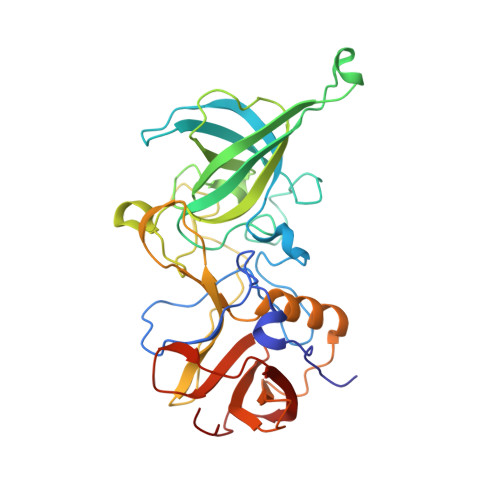
    5HZA, 5HZB

  • PubMed Abstract: 

    Histo-blood group antigens (HBGAs) are important binding factors for norovirus infections. We show that two human milk oligosaccharides, 2'-fucosyllactose (2'FL) and 3-fucosyllactose (3FL), could block norovirus from binding to surrogate HBGA samples. We found that 2'FL and 3FL bound at the equivalent HBGA pockets on the norovirus capsid using X-ray crystallography. Our data revealed that 2'FL and 3FL structurally mimic HBGAs. These results suggest that 2'FL and 3FL might act as naturally occurring decoys in humans.


  • Organizational Affiliation
    • Pediatric Infectious Diseases Unit, University Children's Hospital Mannheim, University of Heidelberg, Mannheim, Germany.

Macromolecule Content 

  • Total Structure Weight: 70.18 kDa 
  • Atom Count: 5,750 
  • Modeled Residue Count: 621 
  • Deposited Residue Count: 630 
  • Unique protein chains: 1

Macromolecules

Find similar proteins by:|  3D Structure
Entity ID: 1
MoleculeChains  Sequence LengthOrganismDetailsImage
Capsid protein
A, B
315Norovirus GII.10Mutation(s): 0 
UniProt
Find proteins for Q5F4T5 (Norwalk virus)
Explore Q5F4T5 
Go to UniProtKB:  Q5F4T5
Entity Groups
Sequence Clusters30% Identity50% Identity70% Identity90% Identity95% Identity100% Identity
UniProt GroupQ5F4T5
Sequence Annotations
Expand
Reference Sequence

Oligosaccharides

Help  
Entity ID: 2
MoleculeChains Length2D Diagram GlycosylationD Interactions
alpha-L-fucopyranose-(1-3)-[beta-D-galactopyranose-(1-4)]beta-D-glucopyranose
C
3N/A
Glycosylation Resources
GlyTouCan: G57548NB
GlyCosmos: G57548NB
GlyGen: G57548NB

Small Molecules

Ligands 1 Unique
IDChains Name / Formula / InChI Key2D Diagram3D Interactions
EDO

Query on EDO


Download:Ideal Coordinates CCD File
D [auth A]
E [auth A]
F [auth A]
G [auth A]
H [auth A]
D [auth A],
E [auth A],
F [auth A],
G [auth A],
H [auth A],
I [auth A],
J [auth B],
K [auth B],
L [auth B],
M [auth B],
N [auth B]
1,2-ETHANEDIOL
C2 H6 O2
LYCAIKOWRPUZTN-UHFFFAOYSA-N

Experimental Data & Validation

Experimental Data

  • Method: X-RAY DIFFRACTION
  • Resolution: 1.35 Å
  • R-Value Free:  0.179 (Depositor), 0.179 (DCC) 
  • R-Value Work:  0.161 (Depositor), 0.161 (DCC) 
  • R-Value Observed: 0.162 (Depositor) 
Space Group: P 1 21 1
Unit Cell:
Length ( Å )Angle ( ˚ )
a = 65.71α = 90
b = 78.17β = 102.3
c = 71.84γ = 90
Software Package:
Software NamePurpose
PHENIXrefinement
XSCALEdata scaling
PHASERphasing
PDB_EXTRACTdata extraction
XDSdata reduction

Structure Validation

View Full Validation Report


Entry History 

Deposition Data

Revision History  (Full details and data files)

  • Version 1.0: 2016-03-02
    Type: Initial release
  • Version 1.1: 2016-04-27
    Changes: Database references
  • Version 2.0: 2020-07-29
    Type: Remediation
    Reason: Carbohydrate remediation
    Changes: Atomic model, Data collection, Derived calculations, Structure summary
  • Version 2.1: 2024-01-10
    Changes: Data collection, Database references, Derived calculations, Refinement description, Structure summary