5HYS

Structure of IgE complexed with omalizumab


Experimental Data Snapshot

  • Method: X-RAY DIFFRACTION
  • Resolution: 2.50 Å
  • R-Value Free: 0.239 
  • R-Value Work: 0.221 
  • R-Value Observed: 0.221 

wwPDB Validation   3D Report Full Report


This is version 2.1 of the entry. See complete history


Literature

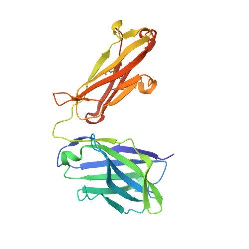
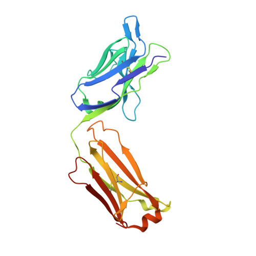
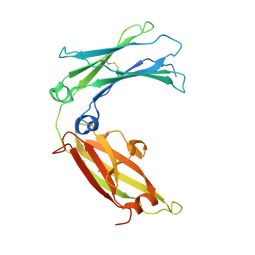
Structural basis of omalizumab therapy and omalizumab-mediated IgE exchange.

Pennington, L.F.Tarchevskaya, S.Brigger, D.Sathiyamoorthy, K.Graham, M.T.Nadeau, K.C.Eggel, A.Jardetzky, T.S.

(2016) Nat Commun 7: 11610-11610

  • DOI: https://doi.org/10.1038/ncomms11610
  • Primary Citation of Related Structures:  
    5HYS

  • PubMed Abstract: 

    Omalizumab is a widely used therapeutic anti-IgE antibody. Here we report the crystal structure of the omalizumab-Fab in complex with an IgE-Fc fragment. This structure reveals the mechanism of omalizumab-mediated inhibition of IgE interactions with both high- and low-affinity IgE receptors, and explains why omalizumab selectively binds free IgE. The structure of the complex also provides mechanistic insight into a class of disruptive IgE inhibitors that accelerate the dissociation of the high-affinity IgE receptor from IgE. We use this structural data to generate a mutant IgE-Fc fragment that is resistant to omalizumab binding. Treatment with this omalizumab-resistant IgE-Fc fragment, in combination with omalizumab, promotes the exchange of cell-bound full-length IgE with omalizumab-resistant IgE-Fc fragments on human basophils. This combination treatment also blocks basophil activation more efficiently than either agent alone, providing a novel approach to probe regulatory mechanisms underlying IgE hypersensitivity with implications for therapeutic interventions.


  • Organizational Affiliation

    Department of Structural Biology, Stanford University School of Medicine, Stanford, California 94305, USA.


Macromolecules
Find similar proteins by:  (by identity cutoff)  |  3D Structure
Entity ID: 1
MoleculeChains Sequence LengthOrganismDetailsImage
Epididymis luminal protein 214A [auth H],
C [auth A],
E [auth C],
G [auth E]
222Homo sapiensMutation(s): 0 
Gene Names: HEL-214
Entity Groups  
Sequence Clusters30% Identity50% Identity70% Identity90% Identity95% Identity100% Identity
Sequence Annotations
Expand
  • Reference Sequence
Find similar proteins by:  (by identity cutoff)  |  3D Structure
Entity ID: 2
MoleculeChains Sequence LengthOrganismDetailsImage
Uncharacterized proteinB [auth L],
D [auth B],
F [auth D],
H [auth F]
218Homo sapiensMutation(s): 0 
Entity Groups  
Sequence Clusters30% Identity50% Identity70% Identity90% Identity95% Identity100% Identity
Sequence Annotations
Expand
  • Reference Sequence
Find similar proteins by:  (by identity cutoff)  |  3D Structure
Entity ID: 3
MoleculeChains Sequence LengthOrganismDetailsImage
Ig epsilon chain C regionI [auth G],
J [auth I],
K [auth J],
L [auth K]
230Homo sapiensMutation(s): 2 
Gene Names: IGHE
UniProt & NIH Common Fund Data Resources
Find proteins for P01854 (Homo sapiens)
Explore P01854 
Go to UniProtKB:  P01854
PHAROS:  P01854
GTEx:  ENSG00000211891 
Entity Groups  
Sequence Clusters30% Identity50% Identity70% Identity90% Identity95% Identity100% Identity
UniProt GroupP01854
Sequence Annotations
Expand
  • Reference Sequence
Oligosaccharides

Help

Entity ID: 4
MoleculeChains Length2D Diagram Glycosylation3D Interactions
alpha-D-mannopyranose-(1-3)-[alpha-D-mannopyranose-(1-6)]beta-D-mannopyranose-(1-4)-2-acetamido-2-deoxy-beta-D-glucopyranose-(1-4)-2-acetamido-2-deoxy-beta-D-glucopyranose
M, N, O, P
5N-Glycosylation
Glycosylation Resources
GlyTouCan:  G22768VO
GlyCosmos:  G22768VO
GlyGen:  G22768VO
Small Molecules
Ligands 1 Unique
IDChains Name / Formula / InChI Key2D Diagram3D Interactions
SO4
Query on SO4

Download Ideal Coordinates CCD File 
AA [auth A]
AB [auth I]
BA [auth B]
BB [auth I]
CA [auth B]
AA [auth A],
AB [auth I],
BA [auth B],
BB [auth I],
CA [auth B],
CB [auth I],
DA [auth B],
DB [auth J],
EA [auth B],
EB [auth J],
FA [auth C],
FB [auth J],
GA [auth C],
GB [auth J],
HA [auth C],
HB [auth K],
IA [auth C],
IB [auth K],
JA [auth D],
JB [auth K],
KA [auth D],
KB [auth K],
LA [auth D],
LB [auth K],
MA [auth D],
NA [auth D],
OA [auth D],
PA [auth E],
Q [auth H],
QA [auth E],
R [auth H],
RA [auth F],
S [auth H],
SA [auth F],
T [auth H],
TA [auth F],
U [auth L],
UA [auth F],
V [auth L],
VA [auth F],
W [auth L],
WA [auth G],
X [auth L],
XA [auth G],
Y [auth A],
YA [auth I],
Z [auth A],
ZA [auth I]
SULFATE ION
O4 S
QAOWNCQODCNURD-UHFFFAOYSA-L
Experimental Data & Validation

Experimental Data

  • Method: X-RAY DIFFRACTION
  • Resolution: 2.50 Å
  • R-Value Free: 0.239 
  • R-Value Work: 0.221 
  • R-Value Observed: 0.221 
  • Space Group: P 1 21 1
Unit Cell:
Length ( Å )Angle ( ˚ )
a = 100.103α = 90
b = 107.143β = 95.18
c = 151.043γ = 90
Software Package:
Software NamePurpose
PHENIXrefinement
HKL-2000data reduction
PHENIXphasing
HKL-2000data scaling

Structure Validation

View Full Validation Report


Entry History & Funding Information

Deposition Data


Funding OrganizationLocationGrant Number
Department of Defense (DOD, United States)United StatesPR130130
National Institutes of Health/National Institute Of Allergy and Infectious Diseases (NIH/NIAID)United StatesU19 AI104209
National Institutes of Health/National Institute Of Allergy and Infectious Diseases (NIH/NIAID)United StatesT32 AI007290

Revision History  (Full details and data files)

  • Version 1.0: 2016-06-01
    Type: Initial release
  • Version 1.1: 2017-09-20
    Changes: Author supporting evidence, Derived calculations
  • Version 1.2: 2019-04-10
    Changes: Author supporting evidence, Data collection
  • Version 1.3: 2019-11-27
    Changes: Author supporting evidence
  • Version 2.0: 2020-07-29
    Type: Remediation
    Reason: Carbohydrate remediation
    Changes: Atomic model, Data collection, Derived calculations, Structure summary
  • Version 2.1: 2023-09-27
    Changes: Data collection, Database references, Derived calculations, Refinement description, Structure summary