5HWA | pdb_00005hwa

Crystal Structure of MH-K1 chitosanase in substrate-bound form


Experimental Data Snapshot

  • Method: X-RAY DIFFRACTION
  • Resolution: 1.35 Å
  • R-Value Free: 
    0.170 (Depositor), 0.165 (DCC) 
  • R-Value Work: 
    0.137 (Depositor) 
  • R-Value Observed: 
    0.138 (Depositor) 

wwPDB Validation 3D Report Full Report

Validation slider image for 5HWA

This is version 2.2 of the entry. See complete history

Literature

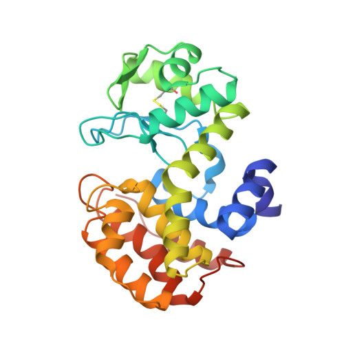
Crystal structure of the GH-46 subclass III chitosanase from Bacillus circulans MH-K1 in complex with chitotetraose

Suzuki, M.Saito, A.Kobayashi, M.Yokoyama, T.Omiya, S.Li, J.Sugita, K.Miki, K.Saito, J.I.Ando, A.

(2024) Biomed Biochim Acta 1868: 130549

Macromolecule Content 

  • Total Structure Weight: 30.42 kDa 
  • Atom Count: 2,681 
  • Modeled Residue Count: 259 
  • Deposited Residue Count: 259 
  • Unique protein chains: 1

Macromolecules

Find similar proteins by:|  3D Structure
Entity ID: 1
MoleculeChains  Sequence LengthOrganismDetailsImage
Chitosanase259Niallia circulansMutation(s): 1 
Gene Names: csn
EC: 3.2.1.132
UniProt
Find proteins for P33673 (Niallia circulans)
Explore P33673 
Go to UniProtKB:  P33673
Entity Groups
Sequence Clusters30% Identity50% Identity70% Identity90% Identity95% Identity100% Identity
UniProt GroupP33673
Sequence Annotations
Expand
Reference Sequence

Oligosaccharides

Help  
Entity ID: 2
MoleculeChains Length2D Diagram GlycosylationD Interactions
2-amino-2-deoxy-beta-D-glucopyranose-(1-4)-2-amino-2-deoxy-beta-D-glucopyranose-(1-4)-2-amino-2-deoxy-beta-D-glucopyranose-(1-4)-2-amino-2-deoxy-beta-D-glucopyranose
B
4N/A
Glycosylation Resources
GlyTouCan: G94774FX
GlyCosmos: G94774FX

Small Molecules

Ligands 3 Unique
IDChains Name / Formula / InChI Key2D Diagram3D Interactions
CAC

Query on CAC


Download:Ideal Coordinates CCD File
C [auth A]CACODYLATE ION
C2 H6 As O2
OGGXGZAMXPVRFZ-UHFFFAOYSA-M
ZN

Query on ZN


Download:Ideal Coordinates CCD File
G [auth A]
H [auth A]
I [auth A]
J [auth A]
K [auth A]
G [auth A],
H [auth A],
I [auth A],
J [auth A],
K [auth A],
L [auth A],
M [auth A]
ZINC ION
Zn
PTFCDOFLOPIGGS-UHFFFAOYSA-N
ACY

Query on ACY


Download:Ideal Coordinates CCD File
D [auth A],
E [auth A],
F [auth A]
ACETIC ACID
C2 H4 O2
QTBSBXVTEAMEQO-UHFFFAOYSA-N

Experimental Data & Validation

Experimental Data

  • Method: X-RAY DIFFRACTION
  • Resolution: 1.35 Å
  • R-Value Free:  0.170 (Depositor), 0.165 (DCC) 
  • R-Value Work:  0.137 (Depositor) 
  • R-Value Observed: 0.138 (Depositor) 
Space Group: P 21 21 21
Unit Cell:
Length ( Å )Angle ( ˚ )
a = 40.75α = 90
b = 79.03β = 90
c = 94.76γ = 90
Software Package:
Software NamePurpose
REFMACrefinement
MOSFLMdata reduction
SCALAdata scaling
MOLREPphasing
PDB_EXTRACTdata extraction

Structure Validation

View Full Validation Report


Entry History 

Deposition Data

Revision History  (Full details and data files)

  • Version 1.0: 2017-02-08
    Type: Initial release
  • Version 1.1: 2020-02-19
    Changes: Data collection
  • Version 2.0: 2020-07-29
    Type: Remediation
    Reason: Carbohydrate remediation
    Changes: Advisory, Atomic model, Data collection, Derived calculations, Structure summary
  • Version 2.1: 2024-02-21
    Changes: Data collection, Database references, Derived calculations, Structure summary
  • Version 2.2: 2024-10-16
    Changes: Data collection, Refinement description, Structure summary