Streptococcus pyogenes quinolinate-salvage pathway-structural and functional studies of quinolinate phosphoribosyl transferase and NH3 -dependent NAD(+) synthetase.
Booth, W.T., Morris, T.L., Mysona, D.P., Shah, M.J., Taylor, L.K., Karlin, T.W., Clary, K., Majorek, K.A., Offermann, L.R., Chruszcz, M.(2017) FEBS J 284: 2425-2441
- PubMed: 28618168 Search on PubMedSearch on PubMed Central
- DOI: https://doi.org/10.1111/febs.14136
- Primary Citation Related Structures:
5HUH, 5HUJ, 5HUL, 5HUO, 5HUP - PubMed Abstract:
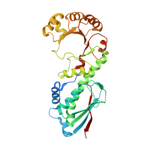
Streptococcus pyogenes, also known as Group A Strep (GAS), is an obligate human pathogen that is responsible for millions of infections and numerous deaths per year. Infection manifestations can range from simple, acute pharyngitis to more complex, necrotizing fasciitis. To date, most treatments for GAS infections involve the use of common antibiotics including tetracycline and clindamycin. Unfortunately, new strains have been identified that are resistant to these drugs, therefore, new targets must be identified to treat drug-resistant strains. This work is focused on the structural and functional characterization of three proteins: spNadC, spNadD, and spNadE. These enzymes are involved in the biosynthesis of nicotinamide adenine dinucleotide (NAD + ). The structures of spNadC and spNadE were determined. SpNadC is suggested to play a role in GAS virulence, while spNadE, functions as an NAD synthetase and is considered to be a new drug target. Determination of the spNadE structure uncovered a putative, NH 3 channel, which may provide insight into the mechanistic details of NH 3 -dependent NAD + synthetases in prokaryotes. Quinolinate phosphoribosyltransferase: EC2.4.2.19 and NAD synthetase: EC6.3.1.5. Protein structures for spNadC, spNadC Δ69A , and spNadE are deposited into Protein Data Bank under the accession codes 5HUL, 5HUO & 5HUP, and 5HUH & 5HUJ, respectively.
- Department of Chemistry and Biochemistry, University of South Carolina, Columbia, SC, USA.
Organizational Affiliation: