5HTK | pdb_00005htk

Human Heart 6-Phosphofructo-2-Kinase/Fructose-2,6-Bisphosphatase (PFKFB2)


Experimental Data Snapshot

  • Method: X-RAY DIFFRACTION
  • Resolution: 2.01 Å
  • R-Value Free: 
    0.175 (Depositor), 0.177 (DCC) 
  • R-Value Work: 
    0.149 (Depositor), 0.153 (DCC) 
  • R-Value Observed: 
    0.151 (Depositor) 

Starting Model: experimental
View more details

wwPDB Validation 3D Report Full Report

Validation slider image for 5HTK

Ligand Structure Quality Assessment 


This is version 1.3 of the entry. See complete history

Literature

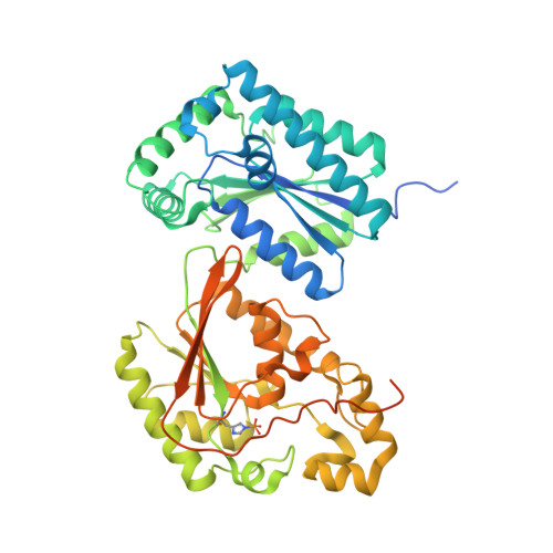
Crystal structure of heart 6-phosphofructo-2-kinase/fructose-2,6-bisphosphatase (PFKFB2) and the inhibitory influence of citrate on substrate binding.

Crochet, R.B.Kim, J.D.Lee, H.Yim, Y.S.Kim, S.G.Neau, D.Lee, Y.H.

(2017) Proteins 85: 117-124

  • DOI: https://doi.org/10.1002/prot.25204
  • Primary Citation Related Structures: 
    5HR5, 5HTK

  • PubMed Abstract: 

    The heart-specific isoform of 6-phosphofructo-2-kinase/fructose-2,6-bisphosphatase (PFKFB2) is an important regulator of glycolytic flux in cardiac cells. Here, we present the crystal structures of two PFKFB2 orthologues, human and bovine, at resolutions of 2.0 and 1.8 Å, respectively. Citrate, a TCA cycle intermediate and well-known inhibitor of PFKFB2, co-crystallized in the 2-kinase domains of both orthologues, occupying the fructose-6-phosphate binding-site and extending into the γ-phosphate binding pocket of ATP. This steric and electrostatic occlusion of the γ-phosphate site by citrate proved highly consequential to the binding of co-complexed ATP analogues. The bovine structure, which co-crystallized with ADP, closely resembled the overall structure of other PFKFB isoforms, with ADP mimicking the catalytic binding mode of ATP. The human structure, on the other hand, co-complexed with AMPPNP, which, unlike ADP, contains a γ-phosphate. The presence of this γ-phosphate made adoption of the catalytic ATP binding mode impossible for AMPPNP, forcing the analogue to bind atypically with concomitant conformational changes to the ATP binding-pocket. Inhibition kinetics were used to validate the structural observations, confirming citrate's inhibition mechanism as competitive for F6P and noncompetitive for ATP. Together, these structural and kinetic data establish a molecular basis for citrate's negative feed-back loop of the glycolytic pathway via PFKFB2. Proteins 2016; 85:117-124. © 2016 Wiley Periodicals, Inc.


  • Organizational Affiliation
    • Departments of Biological Sciences, Louisiana State University, Baton Rouge, Louisiana, 70803.

Macromolecule Content 

  • Total Structure Weight: 120.34 kDa 
  • Atom Count: 7,836 
  • Modeled Residue Count: 845 
  • Deposited Residue Count: 1,010 
  • Unique protein chains: 1

Macromolecules

Find similar proteins by:|  3D Structure
Entity ID: 1
MoleculeChains  Sequence LengthOrganismDetailsImage
6-phosphofructo-2-kinase/fructose-2,6-bisphosphatase 2
A, B
505Homo sapiensMutation(s): 0 
Gene Names: PFKFB2
EC: 2.7.1.105 (PDB Primary Data), 3.1.3.46 (PDB Primary Data)
UniProt & NIH Common Fund Data Resources
Find proteins for O60825 (Homo sapiens)
Explore O60825 
Go to UniProtKB:  O60825
PHAROS:  O60825
GTEx:  ENSG00000123836 
Entity Groups
Sequence Clusters30% Identity50% Identity70% Identity90% Identity95% Identity100% Identity
UniProt GroupO60825
Sequence Annotations
Expand
Reference Sequence

Small Molecules

Ligands 7 Unique
IDChains Name / Formula / InChI Key2D Diagram3D Interactions
ATP

Query on ATP


Download:Ideal Coordinates CCD File
D [auth A],
L [auth B]
ADENOSINE-5'-TRIPHOSPHATE
C10 H16 N5 O13 P3
ZKHQWZAMYRWXGA-KQYNXXCUSA-N
F6P

Query on F6P


Download:Ideal Coordinates CCD File
F [auth A],
N [auth B]
6-O-phosphono-beta-D-fructofuranose
C6 H13 O9 P
BGWGXPAPYGQALX-ARQDHWQXSA-N
MES

Query on MES


Download:Ideal Coordinates CCD File
R [auth B]2-(N-MORPHOLINO)-ETHANESULFONIC ACID
C6 H13 N O4 S
SXGZJKUKBWWHRA-UHFFFAOYSA-N
FLC

Query on FLC


Download:Ideal Coordinates CCD File
C [auth A],
K [auth B]
CITRATE ANION
C6 H5 O7
KRKNYBCHXYNGOX-UHFFFAOYSA-K
GOL

Query on GOL


Download:Ideal Coordinates CCD File
H [auth A]
I [auth A]
J [auth A]
Q [auth B]
S [auth B]
H [auth A],
I [auth A],
J [auth A],
Q [auth B],
S [auth B],
T [auth B],
U [auth B],
V [auth B]
GLYCEROL
C3 H8 O3
PEDCQBHIVMGVHV-UHFFFAOYSA-N
EDO

Query on EDO


Download:Ideal Coordinates CCD File
G [auth A],
O [auth B],
P [auth B]
1,2-ETHANEDIOL
C2 H6 O2
LYCAIKOWRPUZTN-UHFFFAOYSA-N
MG

Query on MG


Download:Ideal Coordinates CCD File
E [auth A],
M [auth B]
MAGNESIUM ION
Mg
JLVVSXFLKOJNIY-UHFFFAOYSA-N
Modified Residues  1 Unique
IDChains TypeFormula2D DiagramParent
NEP
Query on NEP
A, B
L-PEPTIDE LINKINGC6 H10 N3 O5 PHIS

Experimental Data & Validation

Experimental Data

  • Method: X-RAY DIFFRACTION
  • Resolution: 2.01 Å
  • R-Value Free:  0.175 (Depositor), 0.177 (DCC) 
  • R-Value Work:  0.149 (Depositor), 0.153 (DCC) 
  • R-Value Observed: 0.151 (Depositor) 
Space Group: P 21 21 21
Unit Cell:
Length ( Å )Angle ( ˚ )
a = 106.523α = 90
b = 113.946β = 90
c = 133.154γ = 90
Software Package:
Software NamePurpose
PHENIXrefinement
DENZOdata reduction
HKL-2000data scaling

Structure Validation

View Full Validation Report


Ligand Structure Quality Assessment 


Entry History 

Deposition Data

Revision History  (Full details and data files)

  • Version 1.0: 2016-11-16
    Type: Initial release
  • Version 1.1: 2016-12-28
    Changes: Database references
  • Version 1.2: 2020-07-29
    Type: Remediation
    Reason: Carbohydrate remediation
    Changes: Data collection, Derived calculations, Structure summary
  • Version 1.3: 2023-09-27
    Changes: Data collection, Database references, Refinement description, Structure summary