5HSO

Crystal structure of MYCOBACTERIUM TUBERCULOSIS MARR FAMILY PROTEIN Rv2887 complex with DNA


Experimental Data Snapshot

  • Method: X-RAY DIFFRACTION
  • Resolution: 2.50 Å
  • R-Value Free: 0.278 
  • R-Value Work: 0.239 
  • R-Value Observed: 0.241 

wwPDB Validation   3D Report Full Report


This is version 1.2 of the entry. See complete history


Literature

Structural analysis of the regulatory mechanism of MarR protein Rv2887 in M. tuberculosis

Gao, Y.R.Li, D.F.Fleming, J.Zhou, Y.F.Liu, Y.Deng, J.Y.Zhou, L.Zhou, J.Zhu, G.F.Zhang, X.E.Wang, D.C.Bi, L.J.

(2017) Sci Rep 7: 6471-6471

  • DOI: https://doi.org/10.1038/s41598-017-01705-4
  • Primary Citation of Related Structures:  
    5HSM, 5HSO, 5X7Z, 5X80

  • PubMed Abstract: 

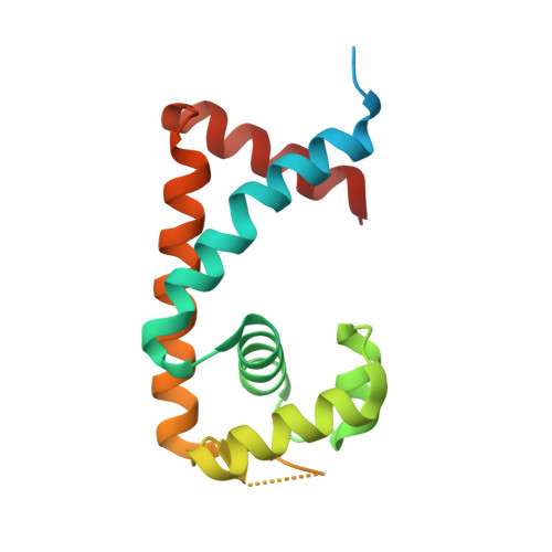
    MarR family proteins are transcriptional regulators that control expression of bacterial proteins involved in metabolism, virulence, stress responses and multi-drug resistance, mainly via ligand-mediated attenuation of DNA binding. Greater understanding of their underlying regulatory mechanism may open up new avenues for the effective treatment of bacterial infections. To gain molecular insight into the mechanism of Rv2887, a MarR family protein in M. tuberculosis, we first showed that it binds salicylate (SA) and para-aminosalicylic acid (PAS), its structural analogue and an antitubercular drug, in a 1:1 stoichiometry with high affinity. Subsequent determination and analysis of Rv2887 crystal structures in apo form, and in complex with SA, PAS and DNA showed that SA and PAS bind to Rv2887 at similar sites, and that Rv2887 interacts with DNA mainly by insertion of helix α4 into the major groove. Ligand binding triggers rotation of the wHTH domain of Rv2887 toward the dimerization domain, causing changes in protein conformation such that it can no longer bind to a 27 bp recognition sequence in the upstream region of gene Rv0560c. The structures provided here lay a foundation for the design of small molecules that target Rv2887, a potential new approach for the development of anti-mycobacterials.


  • Organizational Affiliation

    School of Stomatology and Medicine, Foshan University, Foshan, 528000, Guangdong Province, China.


Macromolecules

Find similar proteins by:  (by identity cutoff)  |  3D Structure
Entity ID: 1
MoleculeChains Sequence LengthOrganismDetailsImage
Uncharacterized HTH-type transcriptional regulator Rv2887
A, B, C, D
160Mycobacterium tuberculosis H37RvMutation(s): 0 
Gene Names: Rv2887MTCY274.18
UniProt
Find proteins for P9WME9 (Mycobacterium tuberculosis (strain ATCC 25618 / H37Rv))
Explore P9WME9 
Go to UniProtKB:  P9WME9
Entity Groups  
Sequence Clusters30% Identity50% Identity70% Identity90% Identity95% Identity100% Identity
UniProt GroupP9WME9
Sequence Annotations
Expand
  • Reference Sequence
Find similar nucleic acids by:  (by identity cutoff)  |  3D Structure
Entity ID: 2
MoleculeChains LengthOrganismImage
DNA (30-MER), the upstream sequence of Rv0560c30Mycobacterium tuberculosis
Sequence Annotations
Expand
  • Reference Sequence
Find similar nucleic acids by:  (by identity cutoff)  |  3D Structure
Entity ID: 3
MoleculeChains LengthOrganismImage
DNA (30-MER), the upstream sequence of Rv0560c30Mycobacterium tuberculosis
Sequence Annotations
Expand
  • Reference Sequence
Experimental Data & Validation

Experimental Data

  • Method: X-RAY DIFFRACTION
  • Resolution: 2.50 Å
  • R-Value Free: 0.278 
  • R-Value Work: 0.239 
  • R-Value Observed: 0.241 
  • Space Group: P 1
Unit Cell:
Length ( Å )Angle ( ˚ )
a = 48.218α = 89.95
b = 48.233β = 90.04
c = 101.115γ = 107.13
Software Package:
Software NamePurpose
PHENIXrefinement
XDSdata reduction
XSCALEdata scaling
PHASERphasing

Structure Validation

View Full Validation Report


Entry History 

Deposition Data

Revision History  (Full details and data files)

  • Version 1.0: 2017-02-01
    Type: Initial release
  • Version 1.1: 2017-08-09
    Changes: Database references
  • Version 1.2: 2024-03-20
    Changes: Data collection, Database references