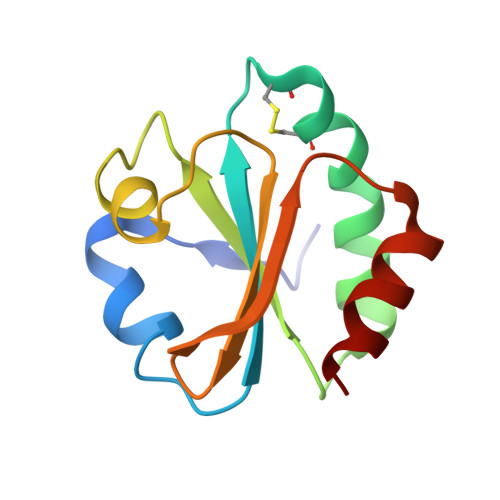
Structural variability of E. coli thioredoxin captured in the crystal structures of single-point mutants.
Noguera, M.E., Vazquez, D.S., Ferrer-Sueta, G., Agudelo, W.A., Howard, E., Rasia, R.M., Manta, B., Cousido-Siah, A., Mitschler, A., Podjarny, A., Santos, J.(2017) Sci Rep 7: 42343-42343
- PubMed: 28181556
- DOI: https://doi.org/10.1038/srep42343
- Primary Citation of Related Structures:
5HR0, 5HR1, 5HR2, 5HR3 - PubMed Abstract:
Thioredoxin is a ubiquitous small protein that catalyzes redox reactions of protein thiols. Additionally, thioredoxin from E. coli (EcTRX) is a widely-used model for structure-function studies. In a previous paper, we characterized several single-point mutants of the C-terminal helix (CTH) that alter global stability of EcTRX. However, spectroscopic signatures and enzymatic activity for some of these mutants were found essentially unaffected. A comprehensive structural characterization at the atomic level of these near-invariant mutants can provide detailed information about structural variability of EcTRX. We address this point through the determination of the crystal structures of four point-mutants, whose mutations occurs within or near the CTH, namely L94A, E101G, N106A and L107A. These structures are mostly unaffected compared with the wild-type variant. Notably, the E101G mutant presents a large region with two alternative traces for the backbone of the same chain. It represents a significant shift in backbone positions. Enzymatic activity measurements and conformational dynamics studies monitored by NMR and molecular dynamic simulations show that E101G mutation results in a small effect in the structural features of the protein. We hypothesize that these alternative conformations represent samples of the native-state ensemble of EcTRX, specifically the magnitude and location of conformational heterogeneity.
- Universidad de Buenos Aires, Facultad de Farmacia y Bioquímica, Instituto de Química y Fisicoquímica Biológicas, CONICET, Junín 956, C1113AAD, Buenos Aires, Argentina.
Organizational Affiliation:


