5HPZ | pdb_00005hpz

type II water soluble Chl binding proteins


Experimental Data Snapshot

  • Method: X-RAY DIFFRACTION
  • Resolution: 1.96 Å
  • R-Value Free: 
    0.266 (Depositor), 0.269 (DCC) 
  • R-Value Work: 
    0.210 (Depositor), 0.217 (DCC) 
  • R-Value Observed: 
    0.213 (Depositor) 

wwPDB Validation 3D Report Full Report

Validation slider image for 5HPZ

Ligand Structure Quality Assessment 


This is version 1.4 of the entry. See complete history

Literature

Fine Tuning of Chlorophyll Spectra by Protein-Induced Ring Deformation.

Bednarczyk, D.Dym, O.Prabahar, V.Peleg, Y.Pike, D.H.Noy, D.

(2016) Angew Chem Int Ed Engl 55: 6901-6905

  • DOI: https://doi.org/10.1002/anie.201512001
  • Primary Citation Related Structures: 
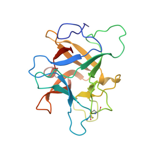
    5HPZ

  • PubMed Abstract: 

    The ability to tune the light-absorption properties of chlorophylls by their protein environment is the key to the robustness and high efficiency of photosynthetic light-harvesting proteins. Unfortunately, the intricacy of the natural complexes makes it very difficult to identify and isolate specific protein-pigment interactions that underlie the spectral-tuning mechanisms. Herein we identify and demonstrate the tuning mechanism of chlorophyll spectra in type II water-soluble chlorophyll binding proteins from Brassicaceae (WSCPs). By comparing the molecular structures of two natural WSCPs we correlate a shift in the chlorophyll red absorption band with deformation of its tetrapyrrole macrocycle that is induced by changing the position of a nearby tryptophan residue. We show by a set of reciprocal point mutations that this change accounts for up to 2/3 of the observed spectral shift between the two natural variants.


  • Organizational Affiliation
    • Department of Biological Chemistry, Weizmann Institute of Science, Rehovot, Israel.

Macromolecule Content 

  • Total Structure Weight: 40.11 kDa 
  • Atom Count: 2,727 
  • Modeled Residue Count: 350 
  • Deposited Residue Count: 358 
  • Unique protein chains: 1

Macromolecules

Find similar proteins by:|  3D Structure
Entity ID: 1
MoleculeChains  Sequence LengthOrganismDetailsImage
Water-soluble chlorophyll protein
A, B
179BrassicaceaeMutation(s): 0 
Gene Names: WSCP1R
UniProt
Find proteins for Q8H0F0 (Brassica oleracea var. viridis)
Explore Q8H0F0 
Go to UniProtKB:  Q8H0F0
Entity Groups
Sequence Clusters30% Identity50% Identity70% Identity90% Identity95% Identity100% Identity
UniProt GroupQ8H0F0
Sequence Annotations
Expand
Reference Sequence

Experimental Data & Validation

Experimental Data

  • Method: X-RAY DIFFRACTION
  • Resolution: 1.96 Å
  • R-Value Free:  0.266 (Depositor), 0.269 (DCC) 
  • R-Value Work:  0.210 (Depositor), 0.217 (DCC) 
  • R-Value Observed: 0.213 (Depositor) 
Space Group: P 2 21 21
Unit Cell:
Length ( Å )Angle ( ˚ )
a = 38.92α = 90
b = 95.479β = 90
c = 98.61γ = 90
Software Package:
Software NamePurpose
REFMACrefinement
MOSFLMdata reduction
PHASERphasing

Structure Validation

View Full Validation Report


Ligand Structure Quality Assessment 


Entry History 

Deposition Data

Revision History  (Full details and data files)

  • Version 1.0: 2016-05-04
    Type: Initial release
  • Version 1.1: 2016-05-18
    Changes: Derived calculations
  • Version 1.2: 2016-06-08
    Changes: Database references
  • Version 1.3: 2019-05-29
    Changes: Data collection, Derived calculations, Structure summary
  • Version 1.4: 2024-10-23
    Changes: Data collection, Database references, Structure summary