5HPN | pdb_00005hpn

A circularly permuted PduA forming an icosahedral cage


Experimental Data Snapshot

  • Method: X-RAY DIFFRACTION
  • Resolution: 2.51 Å
  • R-Value Free: 
    0.256 (Depositor), 0.267 (DCC) 
  • R-Value Work: 
    0.212 (Depositor), 0.226 (DCC) 
  • R-Value Observed: 
    0.216 (Depositor) 

Starting Model: experimental
View more details

wwPDB Validation 3D Report Full Report

Validation slider image for 5HPN

This is version 1.5 of the entry. See complete history

Literature

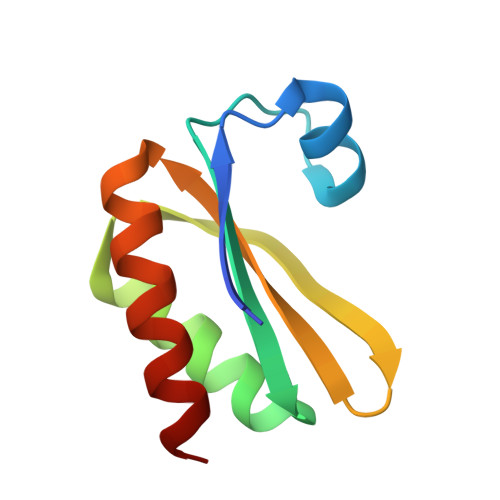
Structure of a novel 13 nm dodecahedral nanocage assembled from a redesigned bacterial microcompartment shell protein.

Jorda, J.Leibly, D.J.Thompson, M.C.Yeates, T.O.

(2016) Chem Commun (Camb) 52: 5041-5044

  • DOI: https://doi.org/10.1039/c6cc00851h
  • Primary Citation Related Structures: 
    5HPN

  • PubMed Abstract: 

    We report the crystal structure of a novel 60-subunit dodecahedral cage that results from self-assembly of a re-engineered version of a natural protein (PduA) from the Pdu microcompartment shell. Biophysical data illustrate the dependence of assembly on solution conditions, opening up new applications in microcompartment studies and nanotechnology.


  • Organizational Affiliation
    • UCLA-DOE Institute for Genomics and Proteomics, University of California, Los Angeles, CA 90095, USA. yeates@mbi.ucla.edu.

Macromolecule Content 

  • Total Structure Weight: 43.99 kDa 
  • Atom Count: 2,890 
  • Modeled Residue Count: 424 
  • Deposited Residue Count: 450 
  • Unique protein chains: 1

Macromolecules

Find similar proteins by:|  3D Structure
Entity ID: 1
MoleculeChains  Sequence LengthOrganismDetailsImage
Permuted PduA
A, B, C, D, E
90synthetic constructMutation(s): 0 

Small Molecules

Ligands 1 Unique
IDChains Name / Formula / InChI Key2D Diagram3D Interactions
SO4

Query on SO4


Download:Ideal Coordinates CCD File
F [auth E]SULFATE ION
O4 S
QAOWNCQODCNURD-UHFFFAOYSA-L

Experimental Data & Validation

Experimental Data

  • Method: X-RAY DIFFRACTION
  • Resolution: 2.51 Å
  • R-Value Free:  0.256 (Depositor), 0.267 (DCC) 
  • R-Value Work:  0.212 (Depositor), 0.226 (DCC) 
  • R-Value Observed: 0.216 (Depositor) 
Space Group: P 42 3 2
Unit Cell:
Length ( Å )Angle ( ˚ )
a = 144.5α = 90
b = 144.5β = 90
c = 144.5γ = 90
Software Package:
Software NamePurpose
PHENIXrefinement
XDSdata reduction
XSCALEdata scaling
PHASERphasing

Structure Validation

View Full Validation Report


Entry History 

& Funding Information

Deposition Data


Funding OrganizationLocationGrant Number
National Science Foundation (NSF, United States)United StatesCHE-1332907

Revision History  (Full details and data files)

  • Version 1.0: 2016-04-06
    Type: Initial release
  • Version 1.1: 2016-04-13
    Changes: Database references
  • Version 1.2: 2016-04-20
    Changes: Derived calculations
  • Version 1.3: 2017-09-20
    Changes: Author supporting evidence, Derived calculations
  • Version 1.4: 2019-11-27
    Changes: Author supporting evidence
  • Version 1.5: 2023-09-27
    Changes: Data collection, Database references, Refinement description