Active Site Desolvation and Thermostability Trade-Offs in the Evolution of Catalytically Diverse Triazine Hydrolases.
Sugrue, E., Carr, P.D., Scott, C., Jackson, C.J.(2016) Biochemistry 55: 6304-6313
- PubMed: 27768291 Search on PubMed
- DOI: https://doi.org/10.1021/acs.biochem.6b00731
- Primary Citation Related Structures:
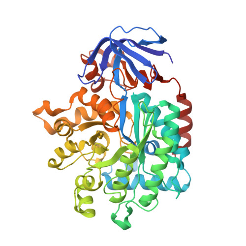
5HMD, 5HME, 5HMF - PubMed Abstract:
The desolvation of ionizable residues in the active sites of enzymes and the subsequent effects on catalysis and thermostability have been studied in model systems, yet little about how enzymes can naturally evolve to include active sites with highly reactive and desolvated charges is known. Variants of triazine hydrolase (TrzN) with significant differences in their active sites have been isolated from different bacterial strains: TrzN from Nocardioides sp. strain MTD22 contains a catalytic glutamate residue (Glu241) that is surrounded by hydrophobic and aromatic second-shell residues (Pro214 and Tyr215), whereas TrzN from Nocardioides sp. strain AN3 has a noncatalytic glutamine residue (Gln241) at an equivalent position, surrounded by hydrophilic residues (Thr214 and His215). To understand how and why these variants have evolved, a series of TrzN mutants were generated and characterized. These results show that desolvation by second-shell residues increases the pK a of Glu241, allowing it to act as a general acid at neutral pH. However, significant thermostability trade-offs are required to incorporate the ionizable Glu241 in the active site and to then enclose it in a hydrophobic microenvironment. Analysis of high-resolution crystal structures shows that there are almost no structural changes to the overall configuration of the active site due to these mutations, suggesting that the changes in activity and thermostability are purely based on the altered electrostatics. The natural evolution of these enzyme isoforms provides a unique system in which to study the fundamental process of charged residue desolvation in enzyme catalysis and its relative contribution to the creation and evolution of an enzyme active site.
- Research School of Chemistry, Australian National University , Canberra, Australia.
Organizational Affiliation: