


Crystal structure of the clinically relevant glutaminase inhibitot CB-839 in complex with glutaminase C
Huang, Q., Katt, W.P., McDermott, L.A., Cerione, R.A.To be published.
Experimental Data Snapshot
Starting Model: experimental
View more details
Entity ID: 1 | |||||
|---|---|---|---|---|---|
| Molecule | Chains | Sequence Length | Organism | Details | Image |
| Glutaminase kidney isoform, mitochondrial | 539 | Homo sapiens | Mutation(s): 0 Gene Names: GLS, GLS1, KIAA0838 EC: 3.5.1.2 | 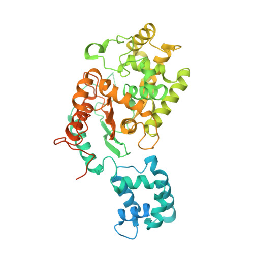 | |
UniProt & NIH Common Fund Data Resources | |||||
GTEx: ENSG00000115419 | |||||
Entity Groups | |||||
| Sequence Clusters | 30% Identity50% Identity70% Identity90% Identity95% Identity100% Identity | ||||
| UniProt Group | O94925 | ||||
Sequence AnnotationsExpand | |||||
Reference Sequence | |||||
| Ligands 1 Unique | |||||
|---|---|---|---|---|---|
| ID | Chains | Name / Formula / InChI Key | 2D Diagram | 3D Interactions | |
| 63J Download:Ideal Coordinates CCD File | E [auth A], F [auth B] | 2-(pyridin-2-yl)-N-(5-{4-[6-({[3-(trifluoromethoxy)phenyl]acetyl}amino)pyridazin-3-yl]butyl}-1,3,4-thiadiazol-2-yl)acetamide C26 H24 F3 N7 O3 S PRAAPINBUWJLGA-UHFFFAOYSA-N |  | ||
| Length ( Å ) | Angle ( ˚ ) |
|---|---|
| a = 98.53 | α = 90 |
| b = 139.382 | β = 90 |
| c = 177.959 | γ = 90 |
| Software Name | Purpose |
|---|---|
| PHENIX | refinement |
| HKL-2000 | data reduction |
| HKL-2000 | data scaling |
| PHASER | phasing |
| Funding Organization | Location | Grant Number |
|---|---|---|
| National Institutes of Health/National Institute of General Medical Sciences (NIH/NIGMS) | United States | GM040654 |
| National Institutes of Health/National Institute of General Medical Sciences (NIH/NIGMS) | United States | GM047458 |


