5HGI | pdb_00005hgi

Crystal structure of apo human IRE1 alpha


Experimental Data Snapshot

  • Method: X-RAY DIFFRACTION
  • Resolution: 2.58 Å
  • R-Value Free: 
    0.247 (Depositor), 0.265 (DCC) 
  • R-Value Work: 
    0.213 (Depositor), 0.225 (DCC) 
  • R-Value Observed: 
    0.214 (Depositor) 

Starting Model: experimental
View more details

wwPDB Validation 3D Report Full Report

Validation slider image for 5HGI

This is version 1.3 of the entry. See complete history

Literature

Structural and Functional Analysis of the Allosteric Inhibition of IRE1 alpha with ATP-Competitive Ligands.

Feldman, H.C.Tong, M.Wang, L.Meza-Acevedo, R.Gobillot, T.A.Lebedev, I.Gliedt, M.J.Hari, S.B.Mitra, A.K.Backes, B.J.Papa, F.R.Seeliger, M.A.Maly, D.J.

(2016) ACS Chem Biol 11: 2195-2205

  • DOI: https://doi.org/10.1021/acschembio.5b00940
  • Primary Citation Related Structures: 
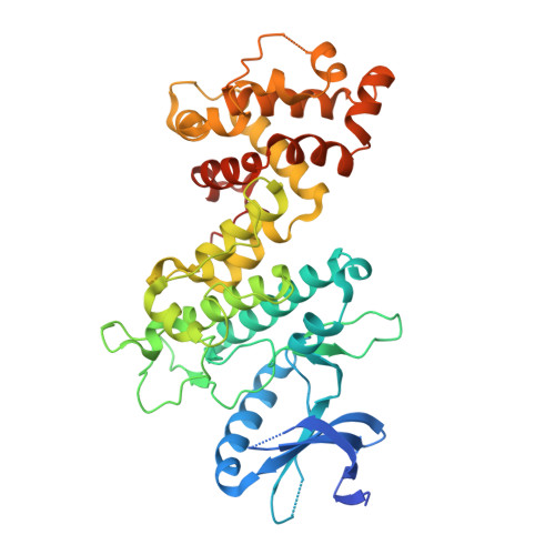
    5HGI

  • PubMed Abstract: 

    The accumulation of unfolded proteins under endoplasmic reticulum (ER) stress leads to the activation of the multidomain protein sensor IRE1α as part of the unfolded protein response (UPR). Clustering of IRE1α lumenal domains in the presence of unfolded proteins promotes kinase trans-autophosphorylation in the cytosol and subsequent RNase domain activation. Interestingly, there is an allosteric relationship between the kinase and RNase domains of IRE1α, which allows ATP-competitive inhibitors to modulate the activity of the RNase domain. Here, we use kinase inhibitors to study how ATP-binding site conformation affects the activity of the RNase domain of IRE1α. We find that diverse ATP-competitive inhibitors of IRE1α promote dimerization and activation of RNase activity despite blocking kinase autophosphorylation. In contrast, a subset of ATP-competitive ligands, which we call KIRAs, allosterically inactivate the RNase domain through the kinase domain by stabilizing monomeric IRE1α. Further insight into how ATP-competitive inhibitors are able to divergently modulate the RNase domain through the kinase domain was gained by obtaining the first structure of apo human IRE1α in the RNase active back-to-back dimer conformation. Comparison of this structure with other existing structures of IRE1α and integration of our extensive structure activity relationship (SAR) data has led us to formulate a model to rationalize how ATP-binding site ligands are able to control the IRE1α oligomeric state and subsequent RNase domain activity.


  • Organizational Affiliation
    • Department of Chemistry, University of Washington , Seattle, Washington, United States.

Macromolecule Content 

  • Total Structure Weight: 50.62 kDa 
  • Atom Count: 3,010 
  • Modeled Residue Count: 395 
  • Deposited Residue Count: 431 
  • Unique protein chains: 1

Macromolecules

Find similar proteins by:|  3D Structure
Entity ID: 1
MoleculeChains  Sequence LengthOrganismDetailsImage
Serine/threonine-protein kinase/endoribonuclease IRE1431Homo sapiensMutation(s): 0 
Gene Names: ERN1IRE1
EC: 2.7.11.1 (PDB Primary Data), 3.1.26 (PDB Primary Data)
UniProt & NIH Common Fund Data Resources
Find proteins for O75460 (Homo sapiens)
Explore O75460 
Go to UniProtKB:  O75460
PHAROS:  O75460
GTEx:  ENSG00000178607 
Entity Groups
Sequence Clusters30% Identity50% Identity70% Identity90% Identity95% Identity100% Identity
UniProt GroupO75460
Sequence Annotations
Expand
Reference Sequence

Small Molecules

Ligands 6 Unique
IDChains Name / Formula / InChI Key2D Diagram3D Interactions
DTT

Query on DTT


Download:Ideal Coordinates CCD File
I [auth A]2,3-DIHYDROXY-1,4-DITHIOBUTANE
C4 H10 O2 S2
VHJLVAABSRFDPM-IMJSIDKUSA-N
CS

Query on CS


Download:Ideal Coordinates CCD File
B [auth A],
C [auth A],
D [auth A],
E [auth A],
F [auth A]
CESIUM ION
Cs
NCMHKCKGHRPLCM-UHFFFAOYSA-N
GOL

Query on GOL


Download:Ideal Coordinates CCD File
H [auth A]GLYCEROL
C3 H8 O3
PEDCQBHIVMGVHV-UHFFFAOYSA-N
BME

Query on BME


Download:Ideal Coordinates CCD File
J [auth A],
K [auth A],
L [auth A],
M [auth A]
BETA-MERCAPTOETHANOL
C2 H6 O S
DGVVWUTYPXICAM-UHFFFAOYSA-N
CL

Query on CL


Download:Ideal Coordinates CCD File
N [auth A],
O [auth A]
CHLORIDE ION
Cl
VEXZGXHMUGYJMC-UHFFFAOYSA-M
NA

Query on NA


Download:Ideal Coordinates CCD File
G [auth A]SODIUM ION
Na
FKNQFGJONOIPTF-UHFFFAOYSA-N

Experimental Data & Validation

Experimental Data

  • Method: X-RAY DIFFRACTION
  • Resolution: 2.58 Å
  • R-Value Free:  0.247 (Depositor), 0.265 (DCC) 
  • R-Value Work:  0.213 (Depositor), 0.225 (DCC) 
  • R-Value Observed: 0.214 (Depositor) 
Space Group: C 2 2 21
Unit Cell:
Length ( Å )Angle ( ˚ )
a = 68.039α = 90
b = 169.196β = 90
c = 104.138γ = 90
Software Package:
Software NamePurpose
PHENIXrefinement
XDSdata reduction
Aimlessdata scaling
PHENIXphasing

Structure Validation

View Full Validation Report


Entry History 

Revision History  (Full details and data files)

  • Version 1.0: 2016-06-08
    Type: Initial release
  • Version 1.1: 2016-06-22
    Changes: Database references
  • Version 1.2: 2016-08-31
    Changes: Database references
  • Version 1.3: 2023-09-27
    Changes: Data collection, Database references, Derived calculations, Refinement description