A Camelid-derived Antibody Fragment Targeting the Active Site of a Serine Protease Balances between Inhibitor and Substrate Behavior
Kromann-Hansen, T., Oldenburg, E., Yung, K.W., Ghassabeh, G.H., Muyldermans, S., Declerck, P.J., Huang, M., Andreasen, P.A., Ngo, J.C.(2016) J Biological Chem 291: 15156-15168
- PubMed: 27226628 Search on PubMedSearch on PubMed Central
- DOI: https://doi.org/10.1074/jbc.M116.732503
- Primary Citation Related Structures:
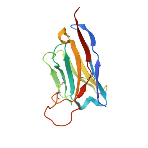
5HDO, 5HGG - PubMed Abstract:
A peptide segment that binds the active site of a serine protease in a substrate-like manner may behave like an inhibitor or a substrate. However, there is sparse information on which factors determine the behavior a particular peptide segment will exhibit. Here, we describe the first x-ray crystal structure of a nanobody in complex with a serine protease. The nanobody displays a new type of interaction between an antibody and a serine protease as it inserts its complementary determining region-H3 loop into the active site of the protease in a substrate-like manner. The unique binding mechanism causes the nanobody to behave as a strong inhibitor as well as a poor substrate. Intriguingly, its substrate behavior is incomplete, as 30-40% of the nanobody remained intact and inhibitory after prolonged incubation with the protease. Biochemical analysis reveals that an intra-loop interaction network within the complementary determining region-H3 of the nanobody balances its inhibitor versus substrate behavior. Collectively, our results unveil molecular factors, which may be a general mechanism to determine the substrate versus inhibitor behavior of other protease inhibitors.
- From the Department of Molecular Biology and Genetics, Aarhus University, Gustav Wieds Vej 10C, 8000 Aarhus C, Denmark, tobiaskh@mbg.au.dk.
Organizational Affiliation: