5HEC | pdb_00005hec

CgT structure in dimer


Experimental Data Snapshot

  • Method: X-RAY DIFFRACTION
  • Resolution: 2.40 Å
  • R-Value Free: 
    0.204 (Depositor), 0.207 (DCC) 
  • R-Value Work: 
    0.163 (Depositor), 0.168 (DCC) 
  • R-Value Observed: 
    0.166 (Depositor) 

Starting Model: experimental
View more details

wwPDB Validation 3D Report Full Report

Validation slider image for 5HEC

This is version 1.5 of the entry. See complete history

Literature

New Helical Binding Domain Mediates a Glycosyltransferase Activity of a Bifunctional Protein.

Zhang, H.Zhou, M.Yang, T.Haslam, S.M.Dell, A.Wu, H.

(2016) J Biological Chem 291: 22106-22117

  • DOI: https://doi.org/10.1074/jbc.M116.731695
  • Primary Citation Related Structures: 
    5HEC

  • PubMed Abstract: 

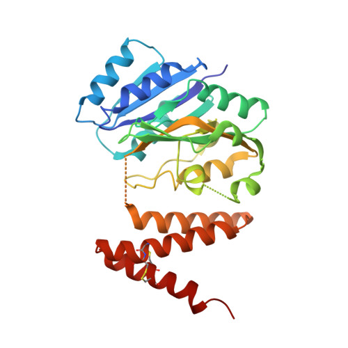
    Serine-rich repeat glycoproteins (SRRPs) conserved in streptococci and staphylococci are important for bacterial colonization and pathogenesis. Fap1, a well studied SRRP is a major surface constituent of Streptococcus parasanguinis and is required for bacterial adhesion and biofilm formation. Biogenesis of Fap1 is a multistep process that involves both glycosylation and secretion. A series of glycosyltransferases catalyze sequential glycosylation of Fap1. We have identified a unique hybrid protein dGT1 (dual glycosyltransferase 1) that contains two distinct domains. N-terminal DUF1792 is a novel GT-D-type glycosyltransferase, transferring Glc residues to Glc-GlcNAc-modified Fap1. C-terminal dGT1 (CgT) is predicted to possess a typical GT-A-type glycosyltransferase, however, the activity remains unknown. In this study, we determine that CgT is a distinct glycosyltransferase, transferring GlcNAc residues to Glc-Glc-GlcNAc-modified Fap1. A 2.4-Å x-ray crystal structure reveals that CgT has a unique binding domain consisting of three α helices in addition to a typical GT-A-type glycosyltransferase domain. The helical domain is crucial for the oligomerization of CgT. Structural and biochemical studies revealed that the helix domain is required for the protein-protein interaction and crucial for the glycosyltransferase activity of CgT in vitro and in vivo As the helix domain presents a novel structural fold, we conclude that CgT represents a new member of GT-A-type glycosyltransferases.


  • Organizational Affiliation
    • From the Departments of Pediatric Dentistry and Microbiology, Schools of Dentistry and Medicine, University of Alabama at Birmingham, Birmingham, Alabama 35294 and.

Macromolecule Content 

  • Total Structure Weight: 68.59 kDa 
  • Atom Count: 4,745 
  • Modeled Residue Count: 552 
  • Deposited Residue Count: 592 
  • Unique protein chains: 1

Macromolecules

Find similar proteins by:|  3D Structure
Entity ID: 1
MoleculeChains  Sequence LengthOrganismDetailsImage
Putative glycosyltransferase (GalT1)
A, B
296Streptococcus parasanguinis FW213Mutation(s): 0 
Gene Names: galT1Spaf_1933
UniProt
Find proteins for I1ZPA1 (Streptococcus parasanguinis FW213)
Explore I1ZPA1 
Go to UniProtKB:  I1ZPA1
Entity Groups
Sequence Clusters30% Identity50% Identity70% Identity90% Identity95% Identity100% Identity
UniProt GroupI1ZPA1
Sequence Annotations
Expand
Reference Sequence

Experimental Data & Validation

Experimental Data

  • Method: X-RAY DIFFRACTION
  • Resolution: 2.40 Å
  • R-Value Free:  0.204 (Depositor), 0.207 (DCC) 
  • R-Value Work:  0.163 (Depositor), 0.168 (DCC) 
  • R-Value Observed: 0.166 (Depositor) 
Space Group: H 3
Unit Cell:
Length ( Å )Angle ( ˚ )
a = 180.044α = 90
b = 180.044β = 90
c = 62.985γ = 120
Software Package:
Software NamePurpose
PHENIXrefinement
HKL-2000data reduction
HKL-2000data scaling
PHENIXphasing

Structure Validation

View Full Validation Report


Entry History 

& Funding Information

Deposition Data


Funding OrganizationLocationGrant Number
National Institutes of Health/National Institute of Dental and Craniofacial Research (NIH/NIDCR)United StatesR01DE017954

Revision History  (Full details and data files)

  • Version 1.0: 2016-08-31
    Type: Initial release
  • Version 1.1: 2016-10-26
    Changes: Database references
  • Version 1.2: 2017-09-27
    Changes: Author supporting evidence, Database references, Derived calculations
  • Version 1.3: 2019-12-11
    Changes: Author supporting evidence
  • Version 1.4: 2023-09-27
    Changes: Data collection, Database references, Refinement description
  • Version 1.5: 2024-10-09
    Changes: Structure summary