5HE2

Bovine GRK2 in complex with Gbetagamma subunits and CCG224406


Experimental Data Snapshot

  • Method: X-RAY DIFFRACTION
  • Resolution: 2.79 Å
  • R-Value Free: 0.277 
  • R-Value Work: 0.198 
  • R-Value Observed: 0.202 

wwPDB Validation   3D Report Full Report


Ligand Structure Quality Assessment 


This is version 1.4 of the entry. See complete history


Literature

Structure-Based Design, Synthesis, and Biological Evaluation of Highly Selective and Potent G Protein-Coupled Receptor Kinase 2 Inhibitors.

Waldschmidt, H.V.Homan, K.T.Cruz-Rodriguez, O.Cato, M.C.Waninger-Saroni, J.Larimore, K.M.Cannavo, A.Song, J.Cheung, J.Y.Kirchhoff, P.D.Koch, W.J.Tesmer, J.J.Larsen, S.D.

(2016) J Med Chem 59: 3793-3807

  • DOI: https://doi.org/10.1021/acs.jmedchem.5b02000
  • Primary Citation of Related Structures:  
    5HE0, 5HE1, 5HE2, 5HE3

  • PubMed Abstract: 

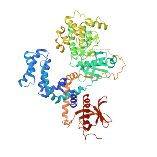
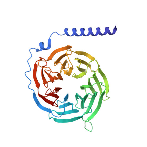
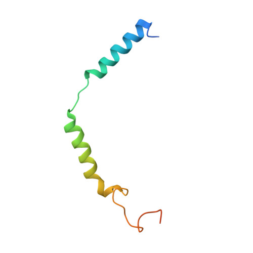
    G protein-coupled receptors (GPCRs) are central to many physiological processes. Regulation of this superfamily of receptors is controlled by GPCR kinases (GRKs), some of which have been implicated in heart failure. GSK180736A, developed as a Rho-associated coiled-coil kinase 1 (ROCK1) inhibitor, was identified as an inhibitor of GRK2 and co-crystallized in the active site. Guided by its binding pose overlaid with the binding pose of a known potent GRK2 inhibitor, Takeda103A, a library of hybrid inhibitors was developed. This campaign produced several compounds possessing high potency and selectivity for GRK2 over other GRK subfamilies, PKA, and ROCK1. The most selective compound, 12n (CCG-224406), had an IC50 for GRK2 of 130 nM, >700-fold selectivity over other GRK subfamilies, and no detectable inhibition of ROCK1. Four of the new inhibitors were crystallized with GRK2 to give molecular insights into the binding and kinase selectivity of this class of inhibitors.


  • Organizational Affiliation

    Center for Translational Medicine, Temple University , Philadelphia, Pennsylvania 19140, United States.


Macromolecules
Find similar proteins by:  (by identity cutoff)  |  3D Structure
Entity ID: 1
MoleculeChains Sequence LengthOrganismDetailsImage
Beta-adrenergic receptor kinase 1641Bos taurusMutation(s): 1 
Gene Names: ADRBK1GRK2
EC: 2.7.11.15
UniProt
Find proteins for P21146 (Bos taurus)
Explore P21146 
Go to UniProtKB:  P21146
Entity Groups  
Sequence Clusters30% Identity50% Identity70% Identity90% Identity95% Identity100% Identity
UniProt GroupP21146
Sequence Annotations
Expand
  • Reference Sequence
Find similar proteins by:  (by identity cutoff)  |  3D Structure
Entity ID: 2
MoleculeChains Sequence LengthOrganismDetailsImage
Guanine nucleotide-binding protein G(I)/G(S)/G(T) subunit beta-1339Homo sapiensMutation(s): 0 
Gene Names: GNB1
UniProt & NIH Common Fund Data Resources
Find proteins for P62873 (Homo sapiens)
Explore P62873 
Go to UniProtKB:  P62873
PHAROS:  P62873
GTEx:  ENSG00000078369 
Entity Groups  
Sequence Clusters30% Identity50% Identity70% Identity90% Identity95% Identity100% Identity
UniProt GroupP62873
Sequence Annotations
Expand
  • Reference Sequence
Find similar proteins by:  (by identity cutoff)  |  3D Structure
Entity ID: 3
MoleculeChains Sequence LengthOrganismDetailsImage
Guanine nucleotide-binding protein G(I)/G(S)/G(O) subunit gamma-2C [auth G]71Homo sapiensMutation(s): 1 
Gene Names: GNG2
UniProt & NIH Common Fund Data Resources
Find proteins for P59768 (Homo sapiens)
Explore P59768 
Go to UniProtKB:  P59768
PHAROS:  P59768
GTEx:  ENSG00000186469 
Entity Groups  
Sequence Clusters30% Identity50% Identity70% Identity90% Identity95% Identity100% Identity
UniProt GroupP59768
Sequence Annotations
Expand
  • Reference Sequence
Small Molecules
Ligands 1 Unique
IDChains Name / Formula / InChI Key2D Diagram3D Interactions
F0S
Query on F0S

Download Ideal Coordinates CCD File 
D [auth A](4~{S})-4-[3-[(2,6-dimethoxyphenyl)methylcarbamoyl]-4-fluoranyl-phenyl]-~{N}-(1~{H}-indazol-5-yl)-6-methyl-2-oxidanylidene-3,4-dihydro-1~{H}-pyrimidine-5-carboxamide
C29 H27 F N6 O5
KQJWWWZXJNOLQF-SANMLTNESA-N
Binding Affinity Annotations 
IDSourceBinding Affinity
F0S BindingDB:  5HE2 IC50: min: 130, max: 130 (nM) from 2 assay(s)
Experimental Data & Validation

Experimental Data

  • Method: X-RAY DIFFRACTION
  • Resolution: 2.79 Å
  • R-Value Free: 0.277 
  • R-Value Work: 0.198 
  • R-Value Observed: 0.202 
  • Space Group: C 2 2 21
Unit Cell:
Length ( Å )Angle ( ˚ )
a = 60.729α = 90
b = 241.95β = 90
c = 213.668γ = 90
Software Package:
Software NamePurpose
REFMACrefinement
HKL-2000data scaling
Cootmodel building
PDB_EXTRACTdata extraction
HKL-2000data reduction
PHASERphasing

Structure Validation

View Full Validation Report


Ligand Structure Quality Assessment 


Entry History & Funding Information

Deposition Data


Funding OrganizationLocationGrant Number
National Institutes of Health/National Heart, Lung, and Blood Institute (NIH/NHLBI)United StatesHL071818
National Institutes of Health/National Heart, Lung, and Blood Institute (NIH/NHLBI)United StatesHL086865
American Heart AssociationUnited StatesN014938
American Heart AssociationUnited States15PRE22730028
Michigan Economic Development Corporation and Michigan Technology Tri-Corridor GrantUnited States085P1000817

Revision History  (Full details and data files)

  • Version 1.0: 2016-05-11
    Type: Initial release
  • Version 1.1: 2017-09-13
    Changes: Author supporting evidence, Database references, Derived calculations
  • Version 1.2: 2017-11-22
    Changes: Refinement description
  • Version 1.3: 2019-12-04
    Changes: Author supporting evidence
  • Version 1.4: 2024-03-06
    Changes: Data collection, Database references